. 2003 Jun 2;22(11):2593-603.
doi: 10.1093/emboj/cdg269.

Crystal structure of tRNA(m1G37)methyltransferase: insights into tRNA recognition

Affiliations

Crystal structure of tRNA(m1G37)methyltransferase: insights into tRNA recognition

Hyung Jun Ahn et al. EMBO J. .

Abstract

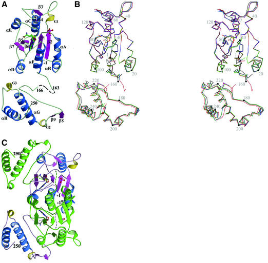
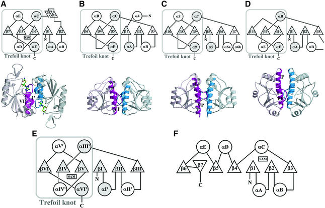
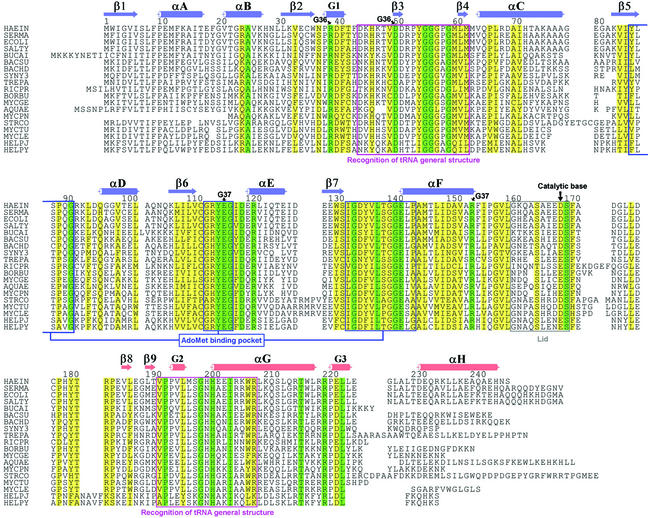
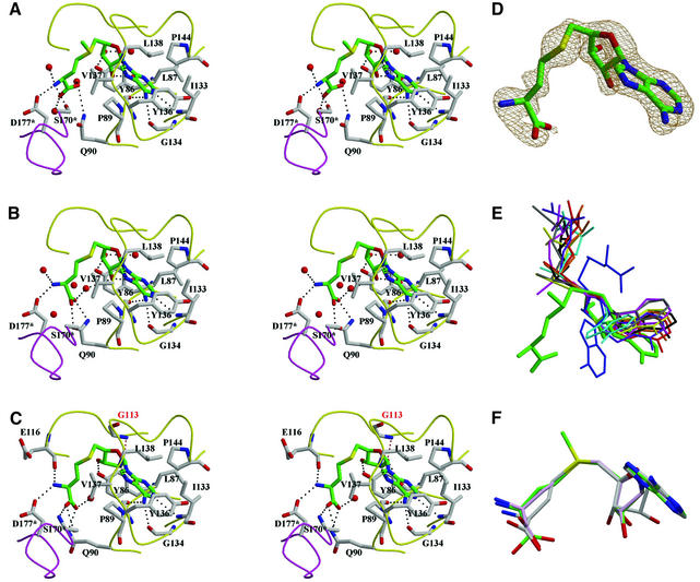
tRNA(m(1)G37)methyltransferase (TrmD) catalyzes the transfer of a methyl group from S-adenosyl-L- methionine (AdoMet) to G(37) within a subset of bacterial tRNA species, which have a G residue at the 36th position. The modified guanosine is adjacent to and 3' of the anticodon and is essential for the maintenance of the correct reading frame during translation. Here we report four crystal structures of TrmD from Haemophilus influenzae, as binary complexes with either AdoMet or S-adenosyl-L-homocysteine (AdoHcy), as a ternary complex with AdoHcy and phosphate, and as an apo form. This first structure of TrmD indicates that it functions as a dimer. It also suggests the binding mode of G(36)G(37) in the active site of TrmD and the catalytic mechanism. The N-terminal domain has a trefoil knot, in which AdoMet or AdoHcy is bound in a novel, bent conformation. The C-terminal domain shows structural similarity to trp repressor. We propose a plausible model for the TrmD(2)-tRNA(2) complex, which provides insights into recognition of the general tRNA structure by TrmD.


Figures

None
Fig. 1. Overall fold of a TrmD monomer. (A) Ribbon diagram (ternary complex). Two missing residues (164 and 165) in the flexible inter-domain linker are shown as dashed lines. G1–G3 represent 310-helices. AdoHcy is shown in a stick model and the phosphate ion in a ball-and-stick model. Carbon atoms are colored in green, oxygen in red, nitrogen in blue, phosphorus in purple, and sulfur in yellow. (B) Stereo view of Cα traces of four models of TrmD. Black is for the apo structure, green for the AdoMet binary complex, blue for the AdoHcy binary complex, and red for the ternary complex with AdoHcy and phosphate. Four models are superimposed in the central five-stranded β-sheet in the NTD. A black dot is marked on every 20th Cα atoms of the apo structure. A comparison of the apo structure and three complex structures shows relatively large r.m.s. differences of 0.66–0.67 Å. The two binary complex structures show the smallest r.m.s. difference of 0.16 Å. R.m.s. differences between binary complex structures and the ternary complex structure are in the intermediate range of 0.39–0.40 Å. (C) Ribbon diagram of a TrmD dimer (ternary complex). One protomer is colored using the same scheme as in (A) and the other protomer is in green. The dashed lines represent the inter-domain linker, which is more ordered in the ternary complex than in binary complexes. Carbon atoms are colored in gray, and other atoms are colored using the same scheme as in (A). This view is obtained by a clockwise rotation of the TrmD monomer in (A) by ∼30° around an axis that is normal to the plane of the figure.
None
Fig. 2. ‘SPOUT class MTase fold’ versus ‘consensus MTase fold’. (A–D) Figures in (A) are for TrmD from H.influenzae, (B) for RmlB from E.coli (PDB entry 1GZ0), (C) for RrmA from T.thermophilus (PDB entry 1IPA), and (D) for MT0001 from M.thermoautotrophicum (PDB entry 1K3R). Figures in the first row are topology diagrams for the ‘SPOUT domain’ of each protein. Common α-helices and β-strands are denoted by circles or triangles filled with gray color. Figures in the second row show the dimer interface between the two ‘SPOUT domains’, each of which comes from different subunits. One monomer is colored in light scarlet and the other in light blue. The two α-helices (αI′ and αVI′) and part of the loop βVI/aVI′ are highlighted in deep colors, and the two α-helices are indicated by Roman numbers, whose definitions are as in (E). (E) Topology diagram of the ‘SPOUT class MTase fold’, as defined in this study. The secondary structure elements that are common to the four SPOUT class MTases are shaded. (F) Topology diagram of the previously defined ‘consensus MTase fold’.
None
Fig. 3. Alignment of 19 TrmD amino acid sequences. Secondary structural elements in the NTD of H.influenzae TrmD are colored in blue, and those in the CTD are colored in red. Strictly conserved residues and highly conserved residues are boxed in green and yellow colors, respectively. The residue numbers are for TrmD from H.influenzae. HAEIN is for TrmD from H.influenzae (SWISS-PROT entry P43912), SERMA for Serratia marcescens (P36244), ECOLI for E.coli (P07020), SALTY for Salmonella typhimurium (P36245), BUCAI for Buchnera aphidicola (P57476), BACSU for Bacillus subtilis (O31741), BACHD for Bacillus halodurans (Q9KA15), SYNY3 for Synechocystis sp. (P72828), TREPA for Treponema pallidum (O83878), RICPR for Rickettsia prowazekii (Q9ZE37), BORBU for Borrelia burgdorferi (O51641), MYCGE for Mycoplasma genitalium (P47683), AQUAE for Aquifex aeolicus (O67463), MYCPN for Mycoplasma pneumoniae (P75132), STRCO for Streptomyces coelicolor (O69882), MYCTU for Mycobacterium tuberculosis (Q10797), MYCLE for Mycobacterium leprae (O33017), HELPJ for Helicobacter pylori J99 (Q9ZK66), and HELPY for Helicobacter pylori 26695 (O25766).
None
Fig. 4. AdoMet/AdoHcy binding mode. (AC) Stereo diagrams of AdoMet in the binary complex and AdoHcy in binary and ternary complexes, respectively. AdoMet/AdoHcy is shown in stick models and colored using the same scheme as in Figure 1A. The residues interacting with AdoMet/AdoHcy are also shown in stick models and their carbon atoms are colored in gray. Water molecules are shown as red balls. Dotted lines represent possible hydrogen bonds between AdoMet/AdoHcy and TrmD or water molecules. The loops colored in yellow are from one protomer and that in purple is from the other protomer. An asterisk after the residue number denotes that the residue comes from the other protomer. In (C), G113 (labeled in red) supports the twist conformation of the ribose ring of AdoHcy through a hydrogen bond, indicated by a red, dotted line. (D) σA-weighted simulated annealing omit map for the binary complex around AdoHcy, contoured at 1.0σ. (E) A novel, bent conformation of AdoMet bound in TrmD (thick lines in green). AdoMet and AdoHcy observed in various MTases are superimposed in the ribose ring. A thin stick model colored in blue represents another distinct bent conformation of AdoHcy in CbiF (PDB entry 1CBF). Extended conformations that are observed most frequently are drawn in thin stick models: AdoMet bound in VP39 (1VPT), ErmC′ (1QAO), FtsJ (1EIZ), M.TaqI (2ADM), M.HhaI (1HMY), PIMT (1JG4) and COMT (1VID) are colored in brown, yellow, red, cyan, black, pink and gray, respectively. AdoHcy bound in CheR (1AF7) is in violet. (F) Superposition of AdoMet and AdoHcy observed in three TrmD complex structures. They are superimposed in the adenine part. Oxygen atoms are colored in red, nitrogen in blue and sulfur in yellow. Carbon atoms in AdoMet are colored in green, those in AdoHcy of the binary complex in light pink, and those in AdoHcy of the ternary complex in gray.
None
Fig. 5. (A) Molecular surface of a TrmD dimer (AdoMet binary complex), excluding a disordered loop covering the active site. Strictly conserved residues and highly conserved residues on the molecular surface are colored in green and yellow, respectively. Two red circles indicate the proposed binding sites for G36 and G37 of the cognate tRNA, respectively. A blue circle indicates the proposed binding site for the hinge region of the L-shaped tRNA. A small black arrow indicates the phosphate-binding position between R24* and R114 in the ternary complex. (B) Proposed catalytic mechanism of H.influenzae TrmD. (C) A proposed model of the TrmD2–tRNA2 complex in the same view as in (A). Each subunit of TrmD, in complex with AdoHcy and phosphate, is colored in blue and scarlet, respectively. AdoHcy is shown in a stick model and colored using the same scheme as in Figure 1A. G36 and G37 of tRNA are highlighted as thick stick models and colored using the same scheme as in Figure 1A. Two other bases of the anti codon are shown in gray stick models. (D) This view is obtained by a clockwise rotation of (C) by 90° around the vertical axis.

References

    1. Anantharaman V, Koonin,E.V. and Aravind,L. (2002) SPOUT: a class of methyltransferases that includes spoU and trmD RNA methylase superfamilies and novel superfamilies of predicted prokaryotic RNA methylases. J. Mol. Microbiol. Biotechnol., 4, 71–75. - PubMed
    1. Batey R.T., Rambo,R.P., Lucast,L., Rha,B. and Doudna,J.A. (2000) Crystal structure of the ribonucleoprotein core of the signal recognition particle. Science, 287, 1232–1239. - PubMed
    1. Battiste J.L., Mao,H., Rao,N.S., Tan,R., Muhandiram,D.R., Kay,L.E., Frankel,A.D. and Williamson,J.R. (1996) α helix–RNA major groove recognition in an HIV-1 Rev peptide–RRE RNA complex. Science, 273, 1547–1551. - PubMed
    1. Björk G.R. (1998) Modified nucleosides in positions 34 and 37 of tRNAs and their predicted coding capacities. In Grosjean,H. and Benne,R. (eds), Modification and Editing of RNA. ASM Press, Washington, DC, pp. 577–581.
    1. Björk G.R., Wikström,P.M. and Byström,A.S. (1989) Prevention of translational frameshifting by the modified nucleoside 1-methyl guanosine. Science, 244, 986–989. - PubMed

Publication types

MeSH terms