. 2003 Jun 2;22(11):2604-13.
doi: 10.1093/emboj/cdg270.

Structures of immature flavivirus particles

Affiliations

Structures of immature flavivirus particles

Ying Zhang et al. EMBO J. .

Abstract

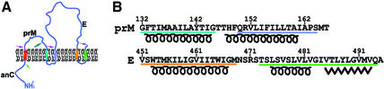
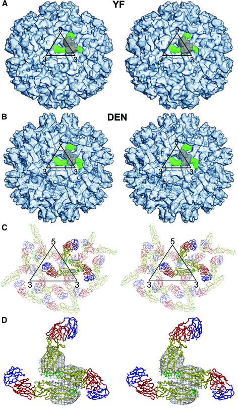
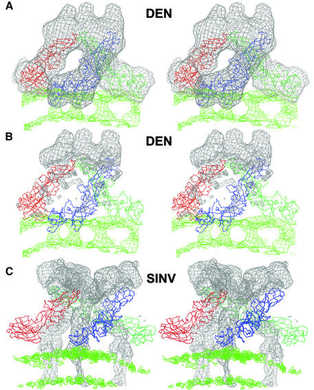
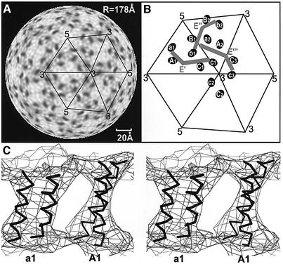
Structures of prM-containing dengue and yellow fever virus particles were determined to 16 and 25 A resolution, respectively, by cryoelectron microscopy and image reconstruction techniques. The closely similar structures show 60 icosahedrally organized trimeric spikes on the particle surface. Each spike consists of three prM:E heterodimers, where E is an envelope glycoprotein and prM is the precursor to the membrane protein M. The pre-peptide components of the prM proteins in each spike cover the fusion peptides at the distal ends of the E glycoproteins in a manner similar to the organization of the glycoproteins in the alphavirus spikes. Each heterodimer is associated with an E and a prM transmembrane density. These transmembrane densities represent either an EE or prMprM antiparallel coiled coil by which each protein spans the membrane twice, leaving the C-terminus of each protein on the exterior of the viral membrane, consistent with the predicted membrane-spanning domains of the unprocessed polyprotein.


Figures

None
Fig. 1. The transmembrane region of dengue polyprotein. (A) The predicted threading of the structural proteins across the membrane. The different transmembrane regions are color coded. Arrows indicate post-translational enzymatic cleavages, with specific enzymes indicated by different colors. The maturation cleavage by furin is shown with a large green arrow (Strauss and Strauss, 2002). (B) Transmembrane amino acid sequences of the prM and E dengue virus proteins color coded to correspond to (A). Secondary structural predictions based on the PROF program (Rost, 1996) are also shown.
None
Fig. 2. Maturation. (A) Maturation of immature prM particle to mature infectious virus and formation of the initial fusogenic particle. (B) The most probable rearrangement required for the immature particles to become mature infectious virions. The Cα backbones of the three independent E molecules per icosahedral asymmetric unit are colored red, blue and green. The three domains in each monomer are identified by Roman numerals: I, central; II, dimerization; III, Ig-like.
None
Fig. 3. Structure of immature flavivirus particles. (A and B) Stereo views showing the surfaces of dengue and yellow fever prM particles at 16 and 25 Å, respectively. An icosahedral asymmetric unit is outlined in black. One of the 60 spikes in each particle is identified in color: prM is gray and E is green. (C) Stereo top view of the organization of the E glycoproteins within and surrounding one icosahedral asymmetric unit (black). Each monomer is shown as a Cα backbone with domains I, II, and III in red, yellow and blue, respectively. The monomers associated with one trimer are in bold. (D) Stereo top view of a spike also showing the density (contoured at a 1σ level) associated with the prM molecule covering the spike structure shown in the same orientation as in (A), (B) and (C). The pairing of the E and prM molecules as a heterodimer is apparent.
None
Fig. 4. Cross-section of prM particle. (A) The ectodomain is shown in blue, the lipid bilayer in green and the nucleocapsid in orange. Plots of maximum height and average height against radius are shown in blue and red, respectively. (B) The equatorial cross-section showing the roughly polygonal shape of the lipid and nucleocapsid. Note the gap between the inner surface of the membrane and the nucleocapsid. This gap is crossed by the nucleocapsid density only at positions corresponding to the base of the external spikes (arrow).
None
Fig. 5. Comparison of the spike structure of immature dengue particles (A and B) and mature Sindbis virus (C). The Cα backbones of the E and E1 glycoprotein trimers (in blue, red and green) are shown for the dengue and Sindbis particles, respectively. (A) The fit of the three E glycoprotein Cα backbones into the spike density (gray) of the immature dengue particle cryoEM density. (B) The density corresponding to the fitted E molecule shown in (A) has been zeroed out, leaving only the density corresponding to the three prM molecules. (C) For comparison, the E1 Cα backbone in the mature Sindbis virus cryoEM density is shown with the density for E1 zeroed out, leaving only the density for E2. The density corresponding to the lipid bilayer is shown in green. The slab used for depicting the lipid bilayer is thinner than that used to define the ectodomain trimers.
None
Fig. 6. The transmembrane structure of the immature particles. (A) Radial section (radius of 178 Å), corresponding roughly to the center of the membrane, showing six elliptical transmembrane regions of higher density arranged in pairs separated by 20 Å. (B) Diagrammatic representation of the transmembrane regions (Aiai, Bibi and Cici, i = 1, 2, 3) and the associated E′, E′′ and E′′′ glycoproteins. The symmetry relationships of E′ to E′′ to E′′′ exactly correspond to the relationships of A1a1 to B2b2 to C3c3, respectively. (C) Stereo view of antiparallel coiled coils for the E and M proteins passing through and back across the membrane at position A1a1. The center of the particle lies below the figure.

References

    1. Allison S.L., Stadler,K., Mandl,C.W., Kunz,C. and Heinz,F.X. (1995) Synthesis and secretion of recombinant tick-borne encephalitis virus protein E in soluble and particulate form. J. Virol., 69, 5816–5820. - PMC - PubMed
    1. Allison S.L., Stiasny,K., Stadler,K., Mandl,C.W. and Heinz,F.X. (1999) Mapping of functional elements in the stem-anchor region of tick-borne encephalitis virus envelope protein E. J. Virol., 73, 5605–5612. - PMC - PubMed
    1. Allison S.L., Schalich,J., Stiasny,K., Mandl,C.W. and Heinz,F.X. (2001) Mutational evidence for an internal fusion peptide in flavivirus envelope protein E. J. Virol., 75, 4268–4275. - PMC - PubMed
    1. Baker T.S. and Cheng,R.H. (1996) A model-based approach for determining orientations of biological macromolecules imaged by cryoelectron microscopy. J. Struct. Biol., 116, 120–130. - PubMed
    1. Baker T.S., Olson,N.H. and Fuller,S.D. (1999) Adding the third dimension to virus life cycles: three-dimensional reconstruction of icosahedral viruses from cryo-electron micrographs. Microbiol. Mol. Biol. Rev., 63, 862–922. - PMC - PubMed

Publication types

MeSH terms