. 2003 Jun 2;22(11):2645-57.
doi: 10.1093/emboj/cdg257.

Rabaptin-5alpha/rabaptin-4 serves as a linker between rab4 and gamma(1)-adaptin in membrane recycling from endosomes

Affiliations

Rabaptin-5alpha/rabaptin-4 serves as a linker between rab4 and gamma(1)-adaptin in membrane recycling from endosomes

Magdalena Deneka et al. EMBO J. .

Abstract

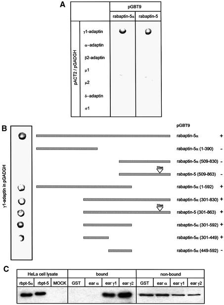
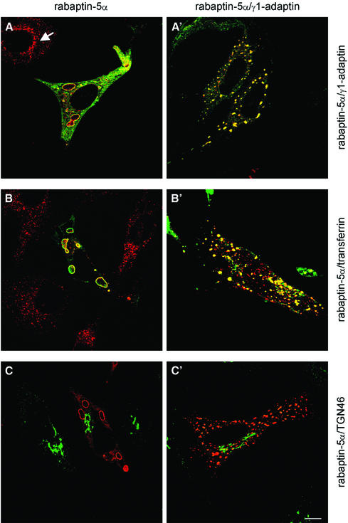
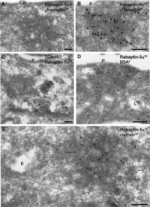
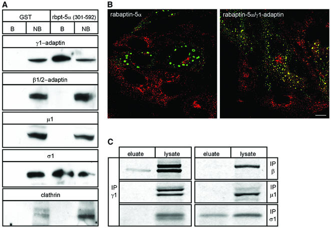
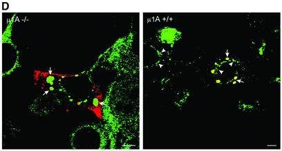
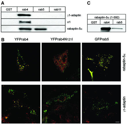
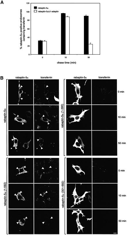
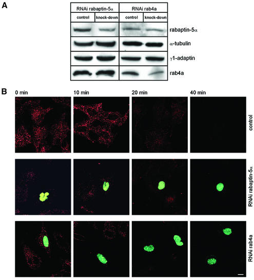
Rab4 regulates recycling from early endosomes. We investigated the role of the rab4 effector rabaptin-5alpha and its putative partner gamma(1)-adaptin in membrane recycling. We found that rabaptin-5alpha forms a ternary complex with the gamma(1)-sigma(1) subcomplex of AP-1, via a direct interaction with the gamma(1)-subunit. The binding site for gamma(1)-adaptin is in the hinge region of rabaptin-5alpha, which is distinct from rab4- and rab5-binding domains. Endogenous or ectopically expressed gamma(1)- adaptin localized to both the trans-Golgi network and endosomes. Co-expressed rabaptin-5alpha and gamma(1)-adaptin, however, co-localized in a rab4-dependent manner on recycling endosomes. Transfection of rabaptin-5alpha caused enlarged endosomes and delayed recycling of transferrin. RNAi of rab4 had an opposing effect on transferrin recycling. Collectively, our data show that rab4-GTP acts as a scaffold for a rabaptin-5alpha- gamma(1)-adaptin complex on recycling endosomes and that interactions between rab4, rabaptin-5alpha and gamma(1)-adaptin regulate membrane recycling.


Figures

None
Fig. 1. Rabaptin-5α(301–449) directly interacts with the ear of γ-adaptin. (A) Yeast two-hybrid assays with rabaptin-5α and rabaptin-5 showing a specific and direct interaction with γ1-adaptin. (B) Summary of two-hybrid assays with rabaptin-5α truncations showing that the domain required for γ1-adaptin binding is between amino acids 301 and 449. Binding is indicated by +. (C) Binding of rabaptin-5α to GST fusions containing the ear domain of γ1-adaptin and γ2-adaptin. GST fusion proteins were incubated with detergent-solubilized brain proteins. Bound proteins were analysed by western blot using a rabaptin-5α antibody. As size control, we used detergent lysates prepared from HeLa cells in which rabaptin-5α and rabaptin-5 were transfected with the vaccinia T7 RNA polymerase system.
None
Fig. 2. Rabaptin-5α–γ-adaptin complex is localized on endosomes. HeLa cells were transfected with rabaptin-5α-pcDNA3.1His (left panels) or with rabaptin-5α and γ1-adaptin-pcDNA3 (right panels). Transfected cells were labelled for rabaptin-5α (green) and γ1-adaptin (red). Note the co-localization of rabaptin-5α and γ1-adaptin in transfected cells, and the distinct localization of γ1-adaptin in non-transfected cells (arrow) (A and A′). Cells were incubated with Alexa594-Tf for 60 min at 37°C and subsequently labelled with anti-Xpress antibody and Alexa488-conjugated IgG (B and B′). The TGN marker TGN46 does not relocate to enlarged endosomes. Transfected cells were labelled for rabaptin-5α (red) and TGN46 (green) (C and C′). Bar, 10 µm.
None
Fig. 3. Rabaptin-5α and γ-adaptin co-localize to tubulo-vesicular membrane clusters. Ultrathin cryosections of HeLa cells transfected with rabaptin- 5α-pcDNA3.1His (A,C and E) or with rabaptin-5α and γ1-adaptin-pcDNA3 (B and D). Double labelling of rabaptin-5α (10 nm gold) and γ1-adaptin (15 nm gold). Rabaptin-5α is associated with clusters of vesicular tubular membranes, where it co-localized with endogenous (A) and overexpressed γ1-adaptin (B). The overall density and diameter of rabaptin-5α-positive membranes is reminiscent of recycling tubules. The dense cytosol between the membranes indicates the presence of high concentrations of cytosolic protein. Note that some of the membranes display a coating typical of the presence of clathrin (arrows in A and B). Double labelling of TGN46 (15 nm gold) and rabaptin-5α (10 nm gold) revealed that rabaptin-5α-positive membranes do not overlap with TGN membranes (C). Rabaptin-5α- (15 nm gold) positive membranes do not contain internalized BSA–5 nm gold. Arrows point to compartments that contain the endocytic marker 10 min after internalization (D). Double labelling of rabaptin-5α (15 nm gold) and clathrin (10 nm gold). Some of the membranes within or associated with rabaptin-5α-positive membranes also stained for clathrin (arrows). Note that the nearby endosomal vacuole (E) has a normal morphology (E). G = Golgi complex, L = lysosome. Bar, 200 nm.
None
Fig. 4. γ1–σ1 AP-1 subcomplex is retained on rabaptin-5α beads. (A) GST or GST–rabaptin-5α(301–592) were incubated with glutathione–Sepharose and a HeLa cell extract. The western blot of bound (B) and non-bound (NB) fractions was probed with antibodies against γ1, β1/2, σ1, µ1 and clathrin. (B) IF of clathrin and rabaptin-5α in cells transfected with rabaptin-5α or co-transfected with rabaptin-5α and γ1-adaptin. Cells were permeabilized, fixed and labelled with a rabbit antibody against rabaptin-5α (green) and X-22 against clathrin (red). (C) HeLa cells were labelled with [35S]methionine and the lysate was incubated with GST–rabaptin-5α(301–592). Bound material was eluted with glutathione. Eluate and cell lysate were incubated with antibodies against γ1 (100/3), β1/2 (100/1), µ1 and σ1, and protein A beads. Immunoprecipitates were resolved by SDS–PAGE and analysed by phosphorimaging. To detect σ1 in the GSH eluate, gels were exposed 10 times longer than for γ1-adaptin (σ1 contains four times less methionine residues than γ1-adaptin). (D) Rabaptin-5α and γ1-adaptin co-localize on endosomes in the absence of intact AP-1. µ1A–/– and µ1A+/+ fibroblasts were transfected with rabaptin-5α-pcDNA3.1His. For γ1-adaptin labelling in µ1A+/+ fibroblasts, cells were extracted with saponin before fixation. Note that γ1-adaptin in these cells is not only present on the structures containing rabaptin-5α (arrows), but also in the TGN area (arrow heads). Saponin treatment of µ1A–/– cells removed all γ1-adaptin labelling (not shown). Cells were fixed, and labelled with a rabbit antibody against rabaptin-5α (red) and mouse γ1-adaptin antibody (Transduction labs) (green). Bar, 10 µm.
None
Fig. 5. Rab4 recruits rabaptin-5α–γ1-adaptin. Rab4, but not rab5 and rab11 interacts with the endogenous rabaptin-5α–γ1–σ1 complex. (A) GST, GST–rab4, GST–rab5 and GST–rab11 were isolated on glutathione–Sepharose, loaded with GTPγS and incubated with pig brain cytosol. Bound fractions were immunoblotted with antibodies against γ1-adaptin, σ1 and rabaptin-5α. (B) Localization of γ1-adaptin to endosomes depends on rab4. HeLa cells were transfected with His-rabaptin-5α in combination with YFP–rab4, dominant-negative YFP–rab4-N121I or GFP–rab5. Cells were labelled with a monoclonal antibody against γ1-adaptin (100/3) (red) and a rabbit antibody against rabaptin-5α (green). Bar, 10 µm. (C) Rab4 is the major binding partner of the N-terminal binding domain on rabaptin-5α. GST fusion proteins were isolated on glutathione–Sepharose, loaded with GTPγS and incubated with 35S-labelled rabaptin-5α(1–592). Bound material was eluted with glutathione, resolved by SDS–PAGE and quantitated by phosphorimaging.
None
Fig. 6. Transfected rabaptin-5α delays Tf recycling. (A) HeLa cells were transfected with rabaptin-5α-pcDNA3.1His or with rabaptin-5α-pcDNA3.1His and γ1-adaptin-pcDNA3. The cells were incubated with 15 µg/ml Alexa-Tf at 16°C for 30 min, and subsequently chased at 37°C. Cells were fixed after different periods of time and stained for rabaptin-5α. Quantitation of the fraction of rabaptin-5α-positive endosomes containing Alexa488-Tf at 0, 10 and 50 min of chase. Error bars denote the standard deviation (n = 10). (B) Rab4 and γ1-adaptin interaction domains in rabaptin-5α are required to retard Tf recycling. HeLa cells were transfected with rabaptin-5α(1–390), rabaptin-5α(301–592), rabaptin-5α(1–592) and rabaptin-5α. The cells were subjected to the pulse–chase protocol as above, fixed and labelled with anti-Xpress followed by Alexa488-labelled anti-mouse IgG. Note that only the rabaptin-5α truncation containing both rab4- and γ1-adaptin-binding sites retarded Tf recycling. Bar, 10 µm.
None
Fig. 7. RNAi of rab4a and rabaptin-5α. (A) HeLa cells were transfected with pKoen (mock), pKoen-rab4a or pKoen-rabaptin-5α for 3 days. Western blots were probed with antibodies against rab4a, rabaptin-5α and tubulin (loading control). In neither case was expression of the other two components of the complex affected. (B) Cells were incubated with 15 µg/ml Alexa-Tf at 16°C for 30 min, chased for different periods of time at 37°C, and processed for fluorescence microscopy. Cells with knocked-down rab4a or rabaptin-5α contain GFP in the nucleus. RNAi of rab4a accelerates exit of Tf from the cells, while reduced rabaptin-5α expression caused a strong inhibition of Tf internalization.

References

    1. Armstrong J.A., Craighead,M.W., Watson,R., Ponnambalam,S. and Bowden,S. (1993) S.pombe ypt5: a homologue of the rab5 endosome fusion regulator. Mol. Biol. Cell, 4, 583–592. - PMC - PubMed
    1. Boehm M. and Bonifacino,J.S. (2001) Adaptins: the final recount. Mol. Biol. Cell, 12, 2907–2920. - PMC - PubMed
    1. Brummelkamp T.R., Bernards,R. and Agami,R. (2002) A system for stable expression of short interfering RNAs in mammalian cells. Science, 296, 550–553. - PubMed
    1. Bucci C., Parton,R., Mather,I., Stunnenberg,H., Simons,K. and Zerial,M. (1992) The small GTPase rab5 functions as a regulatory factor in the early endocytic pathway. Cell, 70, 715–728. - PubMed
    1. Collins B.M., McCoy,A.J., Kent,H.M., Evans,P.R. and Owen,D.J. (2002) Molecular architecture and functional model of the endocytic AP2 complex. Cell, 109, 523–535. - PubMed

Publication types

MeSH terms