

Defective valvulogenesis in HB-EGF and TACE-null mice is associated with aberrant BMP signaling
- PMID: 12773386
- PMCID: PMC156761
- DOI: 10.1093/emboj/cdg264
Defective valvulogenesis in HB-EGF and TACE-null mice is associated with aberrant BMP signaling
Abstract
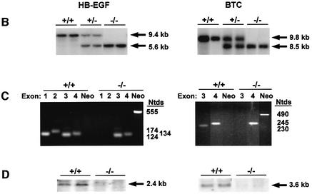
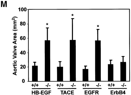
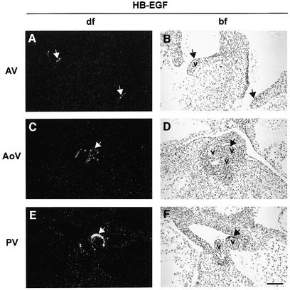
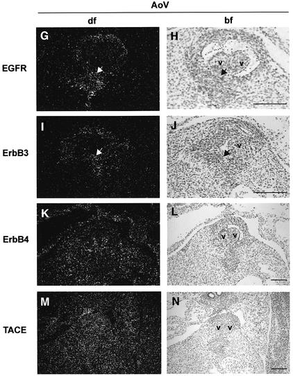
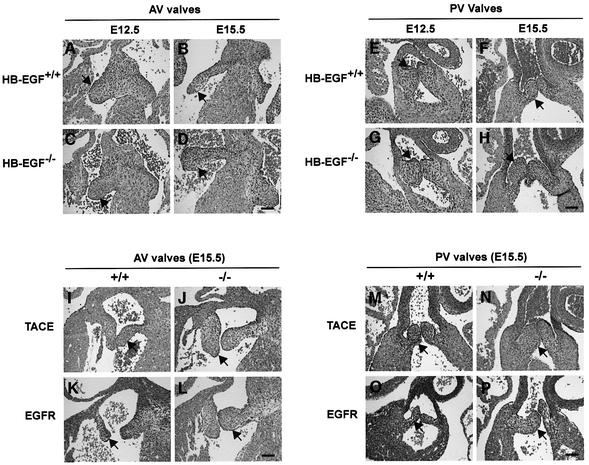
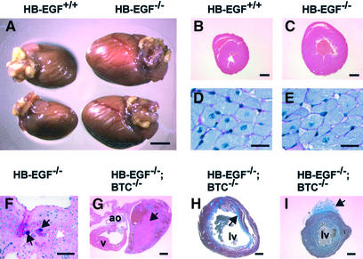
Heparin-binding epidermal growth factor (HB-EGF) and betacellulin (BTC) are activating ligands for EGF receptor (EGFR/ErbB1) and ErbB4. To identify their physiological functions, we disrupted mouse HB-EGF and BTC alleles by homologous recombination. Most HB-EGF(-/-) mice died before weaning, and survivors had enlarged, dysfunctional hearts and reduced lifespans. Although BTC(-/-) mice were viable and fertile and displayed no overt defects, the lifespan of double null HB-EGF(-/-)/BTC(-/-) mice was further reduced, apparently due to accelerated heart failure. HB-EGF(-/-) newborns had enlarged and malformed semilunar and atrioventricular heart valves, and hypoplastic, poorly differentiated lungs. Defective cardiac valvulogenesis was the result of abnormal mesenchymal cell proliferation during remodeling, and was associated with dramatic increases in activated Smad1/5/8. Consistent with the phenotype, HB-EGF transcripts were localized to endocardial cells lining the margins of wild-type valves. Similarly defective valvulogenesis was observed in newborn mice lacking EGFR and tumor necrosis factor-alpha converting enzyme (TACE). These results suggest that cardiac valvulogenesis is dependent on EGFR activation by TACE-derived soluble HB-EGF, and that EGFR signaling is required to regulate bone morphogenetic protein signaling in this context.
Figures


Similar articles
-
Heparin-binding EGF-like growth factor and ErbB signaling is essential for heart function.Proc Natl Acad Sci U S A. 2003 Mar 18;100(6):3221-6. doi: 10.1073/pnas.0537588100. Epub 2003 Mar 5. Proc Natl Acad Sci U S A. 2003. PMID: 12621152 Free PMC article.
-
ErbB1 and ErbB4 generate opposing signals regulating mesenchymal cell proliferation during valvulogenesis.J Cell Sci. 2017 Apr 1;130(7):1321-1332. doi: 10.1242/jcs.196618. Epub 2017 Feb 23. J Cell Sci. 2017. PMID: 28232522
-
Selective roles for tumor necrosis factor alpha-converting enzyme/ADAM17 in the shedding of the epidermal growth factor receptor ligand family: the juxtamembrane stalk determines cleavage efficiency.J Biol Chem. 2004 Jun 4;279(23):24179-88. doi: 10.1074/jbc.M312141200. Epub 2004 Apr 5. J Biol Chem. 2004. PMID: 15066986
-
TACE/ADAM17 processing of EGFR ligands indicates a role as a physiological convertase.Ann N Y Acad Sci. 2003 May;995:22-38. doi: 10.1111/j.1749-6632.2003.tb03207.x. Ann N Y Acad Sci. 2003. PMID: 12814936 Review.
-
ADAM-mediated ectodomain shedding of HB-EGF in receptor cross-talk.Biochim Biophys Acta. 2005 Aug 1;1751(1):110-7. doi: 10.1016/j.bbapap.2004.11.009. Epub 2004 Dec 8. Biochim Biophys Acta. 2005. PMID: 16054021 Review.
Cited by
-
Interleukin-1 stimulates ADAM17 through a mechanism independent of its cytoplasmic domain or phosphorylation at threonine 735.PLoS One. 2012;7(2):e31600. doi: 10.1371/journal.pone.0031600. Epub 2012 Feb 27. PLoS One. 2012. PMID: 22384041 Free PMC article.
-
The phospholipase C isozymes and their regulation.Subcell Biochem. 2012;58:61-94. doi: 10.1007/978-94-007-3012-0_3. Subcell Biochem. 2012. PMID: 22403074 Free PMC article. Review.
-
Dermatitis due to epiregulin deficiency and a critical role of epiregulin in immune-related responses of keratinocyte and macrophage.Proc Natl Acad Sci U S A. 2004 Sep 21;101(38):13921-6. doi: 10.1073/pnas.0404217101. Epub 2004 Sep 13. Proc Natl Acad Sci U S A. 2004. PMID: 15365177 Free PMC article.
-
Signal reachability facilitates characterization of probabilistic signaling networks.BMC Bioinformatics. 2015;16 Suppl 17(Suppl 17):S6. doi: 10.1186/1471-2105-16-S17-S6. Epub 2015 Dec 7. BMC Bioinformatics. 2015. PMID: 26679404 Free PMC article.
-
Stimulation of platelet-derived growth factor receptor beta (PDGFRbeta) activates ADAM17 and promotes metalloproteinase-dependent cross-talk between the PDGFRbeta and epidermal growth factor receptor (EGFR) signaling pathways.J Biol Chem. 2010 Aug 6;285(32):25024-32. doi: 10.1074/jbc.M110.102566. Epub 2010 Jun 7. J Biol Chem. 2010. PMID: 20529858 Free PMC article.
References
-
- Arkonac B.M., Foster,L.C., Sibinga,N.E., Patterson,C., Lai,K., Tsai,J.C., Lee,M.E., Perrella,M.A. and Haber,E. (1998) Vascular endothelial growth factor induces heparin-binding epidermal growth factor-like growth factor in vascular endothelial cells. J. Biol. Chem., 273, 4400–4405. - PubMed
-
- Asakura M. et al. (2002) Cardiac hypertrophy is inhibited by antagonism of ADAM12 processing of HB-EGF: metalloproteinase inhibitors as a new therapy. Nat. Med., 8, 35–40. - PubMed
-
- Bartram U., Bartelings,M.M., Kramer,H.H. and Gittenberger-de Groot, A.C. (2001) Congenital polyvalvular disease: a review. Pediatr. Cardiol., 22, 93–101. - PubMed
-
- Beerli R.R. and Hynes,N.E. (1996) Epidermal growth factor-related peptides activate distinct subsets of ErbB receptors and differ in their biological activities. J. Biol. Chem., 271, 6071–6076. - PubMed
Publication types
MeSH terms
Substances
Grants and funding
LinkOut - more resources
Full Text Sources
Other Literature Sources
Molecular Biology Databases
Research Materials
Miscellaneous
