. 2003 Jun 2;22(11):2741-51.
doi: 10.1093/emboj/cdg271.

The magnitude of hedgehog signaling activity defines skin tumor phenotype

Affiliations

The magnitude of hedgehog signaling activity defines skin tumor phenotype

Vladimir Grachtchouk et al. EMBO J. .

Abstract

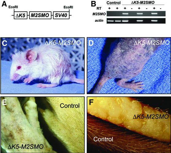
Gain-of-function mutations in SMO have been implicated in constitutive activation of the hedgehog signaling pathway in human basal cell carcinomas (BCCs). We used a truncated keratin 5 (DeltaK5) promoter to assess the potential role of the human M2SMO mutant in BCC development in adult transgenic mice. DeltaK5-M2SMO mouse epidermis is hyperproliferative, ex presses BCC protein markers and gives rise to numerous epithelial downgrowths invading the underlying dermis. Lesions strikingly similar to human basaloid follicular hamartomas develop, but BCCs do not arise even in elderly mice. Hedgehog target gene transcripts were only modestly upregulated in mouse and human follicular hamartomas, in contrast to the high levels detected in BCCs. Cyclins D1 and D2 were selectively upregulated in mouse BCCs. Our data suggest that the levels of hedgehog pathway activation and G(1) cyclins are major determinants of tumor phenotype in skin, and strongly implicate deregulated hedgehog signaling in the genesis of human basaloid follicular hamartomas. Expression of an activated SMO mutant in keratinocytes appears to be insufficient for the development and/or maintenance of full-blown BCCs.


Figures

None
Fig. 1. Transgene design, characterization and gross phenotype of ΔK5-M2SMO mice. (AM2SMO was subcloned into a transgenic cassette containing 1.3 kb of bovine K5 promoter and SV40 small t poly(A) sequence with intron. (B) Expression of M2SMO mRNA in transgenic mouse skin detected by RT–PCR using transgene-specific primers, with β-actin used as an internal control. RT, reverse transcriptase. (C) Three-month-old ΔK5-M2SMO founder with patchy hair loss. (D) An F2 ΔK5-M2SMO601 mouse exhibiting severe phenotype at ∼5 months of age, with generalized alopecia, patchy pigmentation and dry, scaly skin. (E) Close-up of dorsal paw from a ΔK5-M2SMO founder, revealing the haphazard arrangement of the few remaining hair shafts at ∼1 year of age. (F) Extensive alopecia and minute elevations on the surface of the tail from a ΔK5-M2SMO founder at 4.5 months of age, compared with an age-matched control tail.
None
Fig. 2. Altered epidermal growth and differentiation in ΔK5-M2SMO transgenic mice. Epidermal hyperplasia (B) was seen in several ΔK5-M2SMO founders, when compared with age-matched controls (A). epi, epidermis; derm, dermis; hf, hair follicle. Epidermal basal cells in some regions of ΔK5-M2SMO mouse skin were crowded together and elongated (D) in a pattern resembling the peripheral palisading characteristically seen at the edge of human BCC tumor nodules (C). Altered growth and differentiation in skin from ΔK5-M2SMO mice (F, H, J, L, N and P) compared with littermate controls (E, G, I, K, M and O). Note the thickened epidermis and expansion of basal layer-like cells into upper strata (E and F). Expression of K5 is limited to the basal layer in control epidermis (G), but is expressed in multiple suprabasal cell layers in epidermis of ΔK5-M2SMO mice (H). The inset in (G) illustrates normal K5 expression largely limited to the basal layer compartment, even in hyperplastic newborn mouse epidermis. bas, basal cell layer; spin, spinous layers; gran, granular cell layers; cor, cornified cells. The appearance of the suprabasal cell marker K10 is delayed in ΔK5-M2SMO mice (J) compared with controls (I). The inset in (I) shows a normal K10 expression pattern in all suprabasal cell layers of newborn mouse epidermis, but not in the basal cell layer. The dashed line indicates basement membrane separating the epidermis from the dermis. K17 is ectopically expressed in epidermis of ΔK5-M2SMO mice (L) but not controls (K), which exhibit expression limited to the hair follicle outer root sheath. K6 is expressed in the same subset of hair follicle keratinocytes in control (M) and transgenic (N) skin sections. Increased proliferation in ΔK5-M2SMO mouse epidermis (O and P). Note the increased number of BrdU-labeled nuclei (arrowheads) in transgenic (P) versus control (O) mouse skin. BrdU-labeled nuclei and mitotic figures (inset) are seen in suprabasal layers of ΔK5-M2SMO epidermis (P).
None
Fig. 3. Histology, marker expression and increased proliferation rate of basaloid follicular hamartomas in ΔK5-M2SMO mice. H&E-stained skin sections from control (A, C and E) and ΔK5-M2SMO (B, D and F) mice at ∼14 weeks (A–D) and 23 days (E and F) of age. Basaloid follicular hamartomas were detected throughout the integument, including dorsal (A and B) and tail (C–F) skin. In some areas, a cleft separated abnormal epithelium from the surrounding mesenchyme (inset in D), mimicking changes seen in human BCCs. Hair follicles in age-matched control skin (A and C) are in the resting phase of the hair cycle, telogen, with compact clusters of dermal papilla cells (dp) at the tips of the follicles. epi, epidermis; derm, dermis; hf, hair follicle; sq, subcutaneous fat; dp, dermal papilla; seb, sebaceous gland. Analysis of tail skin from young mice (23 days) revealed multiple epithelial downgrowths originating directly from ΔK5-M2SMO epidermis (arrowheads in F, compared with control in E exhibiting a single hair follicle). Note the similar morphology of mouse lesions and human basaloid follicular hamartoma in (G). Marker analysis and BrdU labeling in control (H, J, L and N) and ΔK5-M2SMO transgenic (I, K, M and O) tail skin. Note the widespread expression of K17 in hamartomas (I); expansion of K5 expression (K), which is more evident in immunofluorescence studies (not shown); and focal expression of K10 (M). Scattered pigmented cells frequently are present in the dermis of ΔK5-M2SMO mice (inset in M). Immunostaining for BrdU revealed focally increased proliferation in transgenic mouse skin (O) compared with control (N). (P and Q) Normal growing (anagen) hair follicles. H&E-stained section (P) reveals dermal papilla cells with abundant extracellular material enveloped by hair matrix cells. K17 is expressed in the outer root sheath (Q), but not matrix cells or inner root sheath. ors, outer root sheath; irs, inner root sheath.
None
Fig. 4. Expression of transgene and Shh target genes in follicular hamartomas arising in ΔK5-M2SMO transgenic mice. In situ hybridization of control (A, C and E) and ΔK5-M2SMO skin sections (B, D and F), with epithelial cell islands outlined using dotted lines. Antisense riboprobes hybridizing to the Shh target genes Ptch1 (A and B) and Gli1 (C and D), as well as the transgene-encoded mRNA which contains SV40-derived poly(A) sequence (E and F), revealed patchy expression (blue-purple staining) in a fraction of epithelial cells in follicular hamartomas. Control skin containing resting (telogen) hair follicles did not contain detectable levels of any of these mRNAs. (G) Northern blot analysis revealed elevated expression of Gli1, Gli2 and Ptch1 in RNA samples from ΔK5-M2SMO mouse skin (lanes 1–6), compared with control skin (lanes 7 and 8). (H) Expression of Shh target genes Gli1, Ptch1 and Ptch2, measured by quantitative PCR (TaqMan). RNA was isolated from control skin (n = 3), follicular hamartomas arising in ΔK5-M2SMO mice (n = 6) and BCCs from K5-Gli2 mice (n = 3). Note the dramatic increase in Shh target gene expression in BCCs, with only modest elevations in follicular hamartomas. Standard errors are indicated by bars: the increase in mRNA levels is statistically significant for BCCs (P < 0.05), but not follicular hamartomas.
None
Fig. 5. Distinct skin tumor phenotypes (basaloid follicular hamartoma versus BCC) are associated with different expression levels of Shh target genes and G1 cyclins. (A and B) H&E staining showing basaloid follicular hamartomas, on the left side of each panel, and BCCs, on the right, in sections from K5-Gli2 mice. (CEIn situ analysis reveals abundant expression of Shh target genes Ptch1 and Gli1, and the transgene Gli2, in BCCs on the left side of each panel, but not follicular hamartomas on the right. Negligible expression level of G1 cyclins in control (F and G) and ΔK5-M2SMO (H and I) tail skin. In K5-Gli2 mouse skin, only the BCCs expressed high levels of cyclin D1 (J) and cyclin D2 (K).
None
Fig. 6. Low level expression of SHH target genes in human basaloid follicular hamartomas relative to BCCs. (A and B) In situ analysis for PTCH1 mRNA reveals high expression in essentially all tumor cells in human BCC (A), with low expression levels detected in a fraction of cells comprising basaloid follicular hamartoma (B). (C and D) Low expression of GLI1 mRNA in human follicular hamartoma (D) compared with BCC (C). Note the absence of PTCH1 or GLI1 signal in overlying epidermis of all samples. In situ analysis of five BCCs and five basaloid follicular hamartomas revealed substantially lower PTCH1 and GLI1 expression in all follicular hamartomas.

Similar articles

Cited by

References

    1. Akiyama H., Shigeno,C., Hiraki,Y., Shukunami,C., Kohno,H., Akagi,M., Konishi,J. and Nakamura,T. (1997) Cloning of a mouse smoothened cDNA and expression patterns of hedgehog signalling molecules during chondrogenesis and cartilage differentiation in clonal mouse EC cells, ATDC5. Biochem. Biophys. Res. Commun., 235, 142–147. - PubMed
    1. Aszterbaum M., Epstein,J., Oro,A., Douglas,V., LeBoit,P.E., Scott,M.P. and Epstein,E.H.,Jr (1999) Ultraviolet and ionizing radiation enhance the growth of BCCs and trichoblastomas in patched heterozygous knockout mice. Nat. Med., 5, 1285–1291. - PubMed
    1. Bailleul B., Surani,M.A., White,S., Barton,S.C., Brown,K., Blessing,M., Jorcano,J. and Balmain,A. (1990) Skin hyperkeratosis and papilloma formation in transgenic mice expressing a ras oncogene from a suprabasal keratin promoter. Cell, 62, 697–708. - PubMed
    1. Brown K., Strathdee,D., Bryson,S., Lambie,W. and Balmain,A. (1998) The malignant capacity of skin tumours induced by expression of a mutant H-ras transgene depends on the cell type targeted. Curr. Biol., 8, 516–524. - PubMed
    1. Brownstein M.H. (1992) Basaloid follicular hamartoma: solitary and multiple types. J. Am. Acad. Dermatol., 27, 237–240. - PubMed

Publication types

MeSH terms