. 2003 Jun 2;22(11):2752-63.
doi: 10.1093/emboj/cdg266.

The role of Ppe1/PP6 phosphatase for equal chromosome segregation in fission yeast kinetochore

Affiliations

The role of Ppe1/PP6 phosphatase for equal chromosome segregation in fission yeast kinetochore

Gohta Goshima et al. EMBO J. .

Abstract

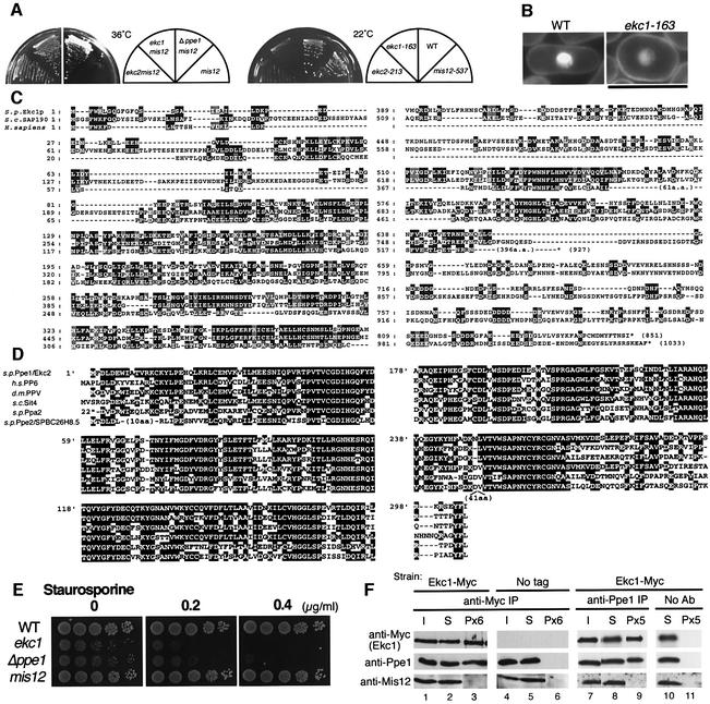
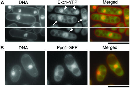
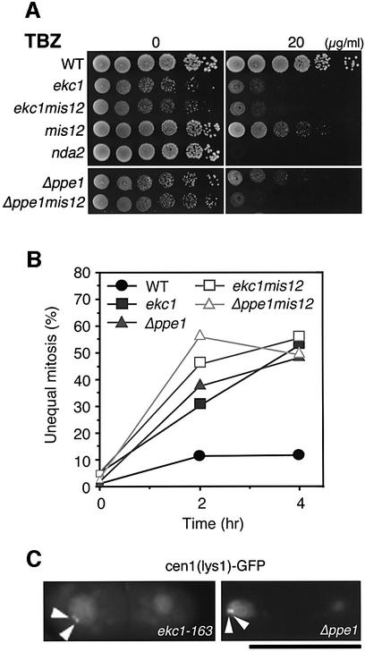
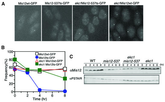
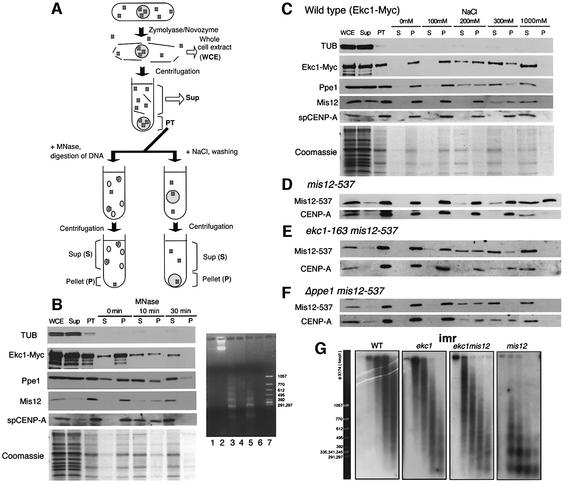
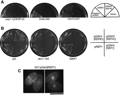
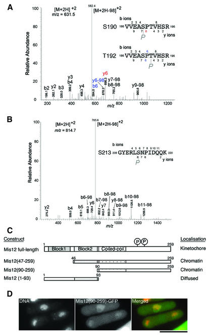
Mis12 is a kinetochore protein essential for equal chromosome segregation and is evolutionarily conserved from yeast to human. In this study, we report the isolation and characterization of suppressors of the mis12 mutant in fission yeast. Our results indicate that Mis12 is negatively regulated by a highly conserved protein phosphatase Ppe1 (scSit4/dmPPV/hPP6) or its bound partner Ekc1 (scSAP), and it is positively regulated by a counteracting kinase Gsk3. Mass spectrometry analysis shows that at least two sites in Mis12 are phosphorylated. This mechanism of suppression occurs at the level of localization recovery of Mis12 to the kinetochore chromatin. Consistently, Mis12 and a subpopulation of Ppe1/Ekc1 were found to behave like non-histone-type chromatin-associating proteins in the chromatin fractionation assay. Mutant analysis of Ppe1 and Ekc1 revealed that they are important for faithful chromosome segregation, as the mutants exhibited unequal chromosome segregation similar to mis12 in the presence of a low concentration of tubulin poison. Ppe1/PP6 directly or indirectly modulates kinetochore chromatin protein Mis12 to ensure progression into normal anaphase.


Figures

None
Fig. 1. Extragenic suppression of mis12-537 by ppe1 and ekc1 mutants. (A) Left: double mutants mis12-537 ekc1-163, mis12-537 ekc2-213 and mis12-537 Δppe1 could form colonies at 36°C, whereas single mis12-537 did not. Right: single ekc1 and ekc2 mutants were cold sensitive and unable to form colonies at 22°C. (Bekc1-163 is pear-shaped even at the permissive temperature. Bar, 10 µm. (C) Amino acid sequence alignment of S.pombe Ekc1, S.cerevisiae SAP190 and Homo sapiens KIAA0685. Identical residues are boxed, and similar residues are hatched. Saccharomyces cerevisiae has four SAPs (SAP4, 190, 185 and 155), while human has three similar sequences (KIAA0685, KIAA1115 and KIAA1558). (D) Sequence alignment of S.pombe Ppe1 phosphatase, S.cerevisiae Sit4, H.sapiens PP6, D.melanogaster PPV and S.pombe Ppa2 and Ppe2/SPBC26H8.05. (E) Exponentially growing wild-type, ekc1, ppe1 and mis12 cells were spotted after dilution onto YPD plate that contained 0.2 or 0.4 µg/ml staurosporine, and incubated at 33°C. (F) The strain containing the integrated Ekc1-Myc was immunoprecipitated by anti-Myc antibodies (lanes 1–3) and anti-Ppe1 antibodies (lanes 7–9), and immunoblotting was used to detect the three proteins Ekc1, Ppe1 and Mis12 in the resulting precipitates. Beads without antibodies (lanes 10 and 11) or the strain without myc epitopes (lanes 4–6) were used as control. I, input; S, supernatant; P, immunoprecipitate. Equal amounts of I and S were loaded, whereas immunoprecipitates were 6- (Px6) or 5-fold (Px5) concentrated.
None
Fig. 2. Nuclear-enriched localization of Ekc1–YFP and Ppe1–GFP. Chromosomally integrated Ekc1–YFP (A, green) and Ppe1–GFP (phosphatase-dead) (B, green) showed nuclear chromatin-enriched localization. 4′,6-diamidino-2-phenylindole (DAPI) was used for DNA staining (red) without fixation. Bars, 10 µm.
None
Fig. 3. Chromosome missegregation in ekc1 and Δppe1 mutants in the presence of TBZ. (A) Moderate hypersensitivity of ekc1-163, Δppe1, mis12, ekc1-163 mis12 and Δppe1 mis12 mutants to TBZ, a microtubule-destabilizing drug. Exponentially growing cells were diluted and spotted onto YPD plates that contained 20 µg/ml TBZ, and incubated at 33°C. nda2-KM52, an α-tubulin mutant, was used as a control hypersensitive mutant. (B) Unequal chromosome segregation frequently occurred in ekc1-163, Δppe1, ekc1 mis12 and Δppe1 mis12 mutant cells in the presence of TBZ. Wild-type and mutant cells were cultured at 33°C, and TBZ (20 µg/ml) was added (time 0). The frequency of dividing cells did not change after TBZ addition, whereas the frequency of mutant cells displaying unequal nuclear division increased to 48–55% (of binucleate cells) after 4 h. (C) Mis-segregation of sister chromatids in ekc1-163 and Δppe1 was confirmed through the use of cen1[lys1]–GFP, a cen1-proximal DNA marker (arrowheads) (33°C, 4 h after TBZ addition). Bar, 10 µm.
None
Fig. 4. Restoration of Mis12 mutant protein localization in the ekc1-163 background. (A) The dot-like localization of Mis12 mutant protein was restored in the double mutant mis12 ekc1. Images represent Mis12–GFP and Mis12-537–GFP expressed in the wild-type and ekc1 background cells (36°C, 8 h). (B) The frequencies of the centromeric dot-like appearance of Mis12ts–GFP were restored in ekc1 mutant cells. Mis12ts–GFP signals in the wild-type background became diffused after 4 h. (CSchizosaccharomyces pombe cell extracts of wild-type and mutant cells cultured at 36°C were prepared at 2 h intervals. Immunoblotting of extracts was performed using anti-Mis12 and control anti-PSTAIR antibodies.
None
Fig. 5. Chromatin fractionation assay of Mis12, CENP-A, Ekc1 and Ppe1 proteins. (A) Schematic representation of the chromatin fractionation assay employed in the present study. See text for explanations. (B) Left: immunoblotting of tubulin (TUB), Ekc1-Myc, Ppe1, Mis12 and spCENP-A in various fractions: WCE, whole-cell extracts; PT, the pellet fraction after spheroplasting; S, supernatant; and P, pellet after MNase treatment for 0, 10 and 30 min. Four-fold concentrated proteins were loaded in the PT, S and P lanes. Mis12, CENP-A, Ekc1-Myc and Ppe1 were found to be solubilized after MNase digestion. Coomassie Blue staining is shown at the bottom. Right: DNA samples extracted before or after MNase digestion (0, 10 and 30 min) were electrophoresed in an agarose gel, followed by ethidium bromide staining. Lane 1, S at 0 min; lane 2, P at 0 min; lane 3, S at 10 min; lane 4, P at 10 min; lane 5, S at 30 min; lane 6, P at 30 min; lane 7, size marker. (C) The PT fraction was washed with the buffer containing 0–1000 mM NaCl followed by centrifugation. Four-fold concentrated proteins were loaded in the PT, S and P lanes. Approximately 30% of Mis12 was solubilized by treatment with 300 mM NaCl. Most Ppe1 and Ekc1-Myc was solubilized in 300 mM NaCl. Coomassie Blue staining is shown at the bottom. (D–F) The same NaCl treatment experiment as (C) was performed for mis12-537 (D), ekc1 mis12-537 (E) and Δppe1 mis12-537 (F) mutants. (G) Recovery of centromere chromatin structure by ekc1 mutation. Nuclear chromatin was prepared from spheroplasts of wild type and mis12, ekc1 and ekc1 mis12 mutants cultured at 36°C for 8 h. Chromatin was digested with MNase for 0, 1, 2, 4 and 8 min (left to right lanes). Digested DNAs were electrophoresed in an agarose gel. Southern hybridization using the central centromere DNA probe imr1 was performed. The size markers are shown on the left.
None
Fig. 6. Overproduced Gsk3 kinase suppresses mis12 and antagonizes Ppe1. (A) Multicopy plasmid pGsk3 suppressed the ts phenotype of mis12-537 at 36°C, but did not suppress cnp1-1 and mis6-302 mutants even at 33°C. (B) Gsk3 is antagonistic to Ppe1 and Ekc1, as its moderate overexpression by REP41 plasmid in the absence of thiamine prevented colony formation of ekc1-163 and Δppe1, but not of wild-type. Even the milder expression of Gsk3 by REP81 plasmid still caused retarded growth of ekc1 and Δppe1 mutants. REP1 caused the strongest overexpression. (C) Gsk3 overexpression in the wild-type cells led to the accumulation of pear-shaped cells. Wild-type cells in which Gsk3 was expressed by REP1 plasmid in the absence of thiamine for 24 h at 26°C. Cells were stained by DAPI. Bar, 10 µm.
None
Fig. 7. Residues in the C-terminus of Mis12 are phosphorylated. (A and B) Ser190 or Thr192 and Ser213 are phosphorylated in vivo. HA- and histidine-tagged Mis12 overproduced in wild type was purified using TALON beads under denaturing conditions, and subjected to SDS–PAGE. The band of Mis12-HAHis6 was cut out and trypsinized in-gel, and subjected to survey of the phosphorylation site using MS (Ohta et al., 2002). The MS/MS spectra of the tryptic phosphopeptide amino acids 186–196 (A) and 208–220 (B) of Mis12 obtained by collision-induced dissociation of the [M + 2H]2+precursor ions, m/z 631.5 and 814.7, are shown. In both cases, intense [M + 2H –98]2+ ions, m/z 582.4 in (A) and 765.8 in (B), due to neutral loss (H2O + phosphate, 97.8) of precursor ions were observed, indicating that the potential phosphorylated residues are serine or threonine. Three potential sites of phosphorylation (two serine and one threonine) are found in the peptide 186–196 (A). The fragment ions enable localization of the phosphorylated residue to one of the two central residues, Ser190 and Thr192. An ion fragment at m/z 696.3 is assignable to y6 (red letters) when the peptide is phosphorylated at Ser190, and ion fragments at m/z 584.0 and 678.3 are assignable to b6 and y6-98, respectively, when the peptide is phosphorylated at Thr192 (blue letters). There is one possible residue in the peptide 208–220 (B), and the fragment ions were matched to the peptide phosphorylated at Ser213. (C) Localization of three Mis12-truncated proteins. Each construct is tagged with GFP at the C-terminus and expressed from REP41 plasmid that allows moderate overexpression. (D) Chromatin localization of truncated Mis12 (amino acids 90–259)–GFP protein. DNA was counterstained by DAPI. Bar, 10 µm.

Similar articles

Cited by

References

    1. Bastians H. and Ponstingl,H. (1996) The novel human protein serine/threonine phosphatase 6 is a functional homologue of budding yeast Sit4p and fission yeast ppe1, which are involved in cell cycle regulation. J. Cell Sci., 109, 2865–2874. - PubMed
    1. Beck T. and Hall,M.N. (1999) The TOR signalling pathway controls nuclear localization of nutrient-regulated transcription factors. Nature, 402, 689–692. - PubMed
    1. Biggins S., Severin,F.F., Bhalla,N., Sassoon,I., Hyman,A.A. and Murray,A.W. (1999) The conserved protein kinase Ipl1 regulates microtubule binding to kinetochores in budding yeast. Genes Dev., 13, 532–544. - PMC - PubMed
    1. Blower M.D. and Karpen,G.H. (2001) The role of Drosophila CID in kinetochore formation, cell-cycle progression and heterochromatin interactions. Nat. Cell Biol., 3, 730–739. - PMC - PubMed
    1. Cohen P.T. (1997) Novel protein serine/threonine phosphatases: variety is the spice of life. Trends Biochem. Sci., 22, 245–251. - PubMed

Publication types

MeSH terms