. 2003 Jun;23(12):4056-65.
doi: 10.1128/MCB.23.12.4056-4065.2003.

Methylation of adjacent CpG sites affects Sp1/Sp3 binding and activity in the p21(Cip1) promoter

Affiliations

Methylation of adjacent CpG sites affects Sp1/Sp3 binding and activity in the p21(Cip1) promoter

Wei-Guo Zhu et al. Mol Cell Biol. 2003 Jun.

Abstract

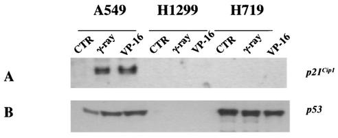
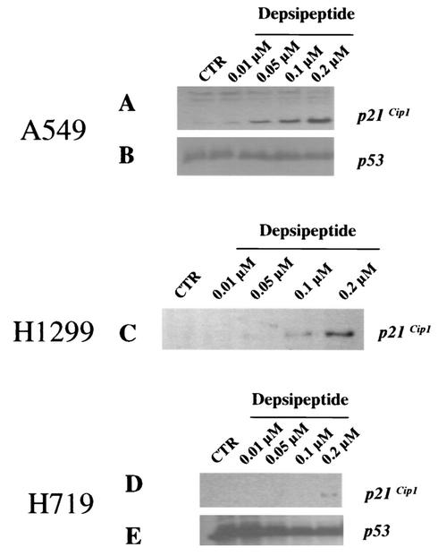
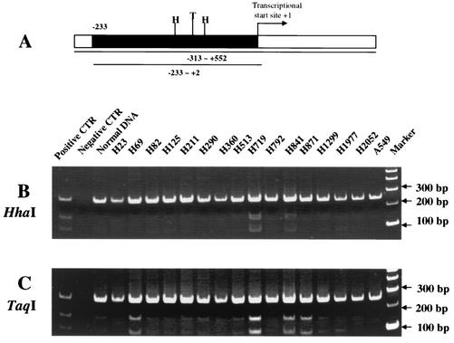
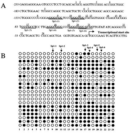
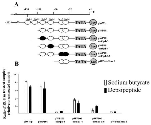
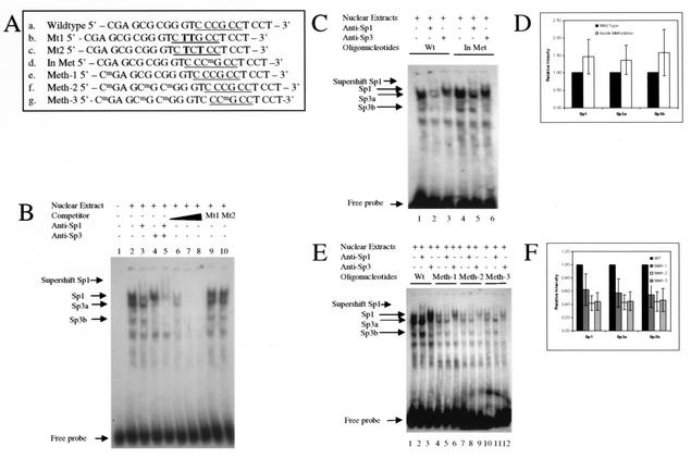
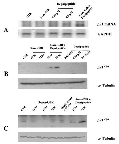
DNA methylation in the promoter of certain genes is associated with transcriptional silencing. Methylation affects gene expression directly by interfering with transcription factor binding and/or indirectly by recruiting histone deacetylases through methyl-DNA-binding proteins. In this study, we demonstrate that the human lung cancer cell line H719 lacks p53-dependent and -independent p21(Cip1) expression. p53 response to treatment with gamma irradiation or etoposide is lost due to a mutation at codon 242 of p53 (C-->W). Treatment with depsipeptide, an inhibitor of histone deacetylase, was unable to induce p53-independent p21(Cip1) expression because the promoter of p21(Cip1) in these cells is hypermethylated. By analyzing luciferase activity of transfected p21(Cip1) promoter vectors, we demonstrate that depsipeptide functions on Sp1-binding sites to induce p21(Cip1) expression. We hypothesize that hypermethylation may interfere with Sp1/Sp3 binding. By using an electrophoretic mobility shift assay, we show that, although methylation within the consensus Sp1-binding site did not reduce Sp1/Sp3 binding, methylation outside of the consensus Sp1 element induced a significant decrease in Sp1/Sp3 binding. Depsipeptide induced p21(Cip1) expression was reconstituted when cells were pretreated with 5-aza-2'-deoxycytidine. Our data suggest, for the first time, that hypermethylation around the consensus Sp1-binding sites may directly reduce Sp1/Sp3 binding, therefore leading to a reduced p21(Cip1) expression in response to depsipeptide treatment.


Figures

FIG. 1.
FIG. 1.
Expression of p21Cip1 and p53 in human lung cancer cells in response to gamma irradiation or etoposide treatment. Cells were exposed to gamma irradiation (8 Gy) or VP-16 treatment (50 μg/ml for 24 h) and then incubated at 37°C for 24 h. Protein was harvested, and Western immunoblot was performed to detect the expression of p21Cip1 (A) and p53 (B).
FIG. 2.
FIG. 2.
Expression of p21Cip1 and p53 in human lung cancer cells after depsipeptide treatment. Cells were treated with depsipeptide (0.0125 to 0.2 μM for 6 h), washed with cold PBS, and incubated at 37°C for 24 h. Protein was harvested, and Western immunoblot analysis was performed to detect the expression of p21Cip1 (A, C, and D) or p53 (B and E) in response to depsipeptide treatment. H1299 cells have no endogenous p53.
FIG. 3.
FIG. 3.
Methylation status of p21Cip1 promoter in human lung cancer cells. (A) Schematic diagram of the p21Cip1 promoter. The CpG island of p21Cip1 promoter (positions −313 to +552 relative to the transcription start site; National Center for Biotechnology Information [NCBI] no. U24170) was determined by computer program (WebGene [http://www.itba.mi.cnr.it/webgene/]). Primers were designed to amplify a fragment spanning positions −233 to +2 (235 bp, black box). Within this fragment there is one TaqI recognition site (TCGA, presented as T in the figure) and two HhaI recognition sites (GCGC, presented as H in the figure). (B and C) DNA from 16 human lung cancer cell lines and normal human blood cells was treated with bisulfite and then PCR amplified. The PCR products were purified and digested with the CG-containing enzymes HhaI (B) or TaqI (C). Digested samples were size separated by 8% PAGE. DNA not treated with bisulfite served as a negative control. DNA treated with Sss methylase was the positive control. Digested fragments correspond to methylated DNA.
FIG. 4.
FIG. 4.
Bisulfite sequencing of CpG island in the p21Cip1 promoter (A) The CpG island of p21Cip1 promoter (NCBI no. U24170) was analyzed. This sequence spans 250 bp between positions −233 to +17 relative to the transcription start site, including 24 CGs upstream of the transcriptional start site. There are six CG-containing Sp1-binding sites within this sequence that are indicated as underlined and emboldened, corresponding to the 9th, 11th, 17th, 18th, 20th, and 21st CGs within this island. (B) DNA from H719 cells was treated with bisulfite, and the p21Cip1 promoter was PCR amplified. The PCR product was ligated into pCR2.1-TOPO by using the TA cloning system. Twenty subclones were picked and sequenced. Symbols: ○, unmethylated cytosines; •, methylated cytosines.
FIG. 5.
FIG. 5.
Analysis of relative luciferase activity in cells treated with depsipeptide or sodium butyrate. (A) p21Cip1 promoter constructs used for the luciferase transfection assay. The human wild-type p21Cip1 promoter luciferase fusion plasmid, pWWP-Luc, contains all six Sp1 sites and the transcription start site (2.4 kb). pWP101 contains four Sp1 sites termed Sp1-3 to Sp1-6. pWPdel-SmaI only contains the Sp1-5 and Sp1-6 sites. Three mutated Sp1 vectors are pWP101mtSp1-3 (mutated at the Sp1-3 site), pWP101mtSp1-4 (mutated at the Sp1-4 site), and pWP101mtSp1-5,6 (mutated at the Sp1-5 and Sp1-6 sites). •, mutated Sp1-binding sites. (B) At 24 h after transfection, H719 cells were treated with 0.05 μM depsipeptide for 6 h or 5 mM sodium butyrate for 24 h. The cells were harvested for analysis of luciferase activity at 24 h after the treatments. The luciferase activity of each sample was normalized for the amount of protein in the cell lysate. The experiments were carried out at least two times in triplicate. The luciferase activity of the untreated cells that was transfected with wild-type pWWP-Luc vector served as a control. The relative luciferase activity for each sample was then compared to the control, and the ratios were calculated.
FIG. 6.
FIG. 6.
Analysis of binding of Sp1/Sp3 to the recognition Sp1-3-binding sites assayed by EMSA. (A) Sequence of oligonucleotides used for EMSA experiments (positions −93 to −72 relative to transcription start site of the p21Cip1 promoter). Sequences: a, wild-type Sp1-3-binding site of p21Cip1 promoter (Sp1-binding site is underlined); b and c, mutated Sp1-3-binding sites, with the mutated nucleotides in boldface; d, methylated Sp1-3-binding site within the Sp1-3-binding site (the methylated C corresponds to CG site 17 in Fig. 4); e and f, methylated cytosines located upstream of the consensus Sp1-3-binding site (the methylated C's correspond to CG sites 14, 15, and 16 in Fig. 4); g, methylated cytosines located both in and upstream of the Sp1-3-binding site. (B) EMSA experiments show specific binding for Sp1 and Sp3 (from H719 nuclear extracts) to the Sp1-3 recognition binding site. Lane 1, radiolabeled probe in the absence of nuclear extract; lane 2, Sp1/Sp3 binding is depicted by arrows on left of figures; lane 3, anti-Sp1 antibody is added to extracts; lane 4, anti-Sp3 supershift; lane 5, anti-Sp1 and anti-Sp3 supershift; lanes 6 to 8, excess unlabeled wild-type competitor (lane 6 [1:10], lane 7 [1:50], and lane 8 [1:100]) competes for binding with the labeled element; lanes 9 and 10, unlabeled mutated Sp1-3 oligonucleotide (1:100) could not compete with the labeled Sp1 probe. (C) Comparison of nuclear extract binding to the consensus wild-type and methylated Sp1-3-binding sites (oligonucleotides a [wild type] and d [In Met], respectively). Lanes 1 to 3 show binding reactions with labeled wild-type Sp1-3 as a probe, and lanes 4 to 6 show binding reactions with labeled methylated Sp1-3 as a probe. Lanes 1 and 4, no antibody added; lanes 2 and 5, anti-Sp1 antibody; lanes 3 and 6, anti-Sp3 antibody. (D) Graphic representation of the the relative binding affinities of nuclear extracts to the oligonucleotides in panel C (lanes 1 and 4). The relative intensity of binding was determined in four replicate experiments. (E) Comparison of nuclear extract binding to the consensus Sp1-3-binding element with wild-type and nonconsensus methylated oligonucleotides (see panel A, oligonucleotides e to g). Lanes 1 to 3, wild-type Sp1-3 element; lanes 4 to 6, oligonucleotide e (Meth-1); lanes 7 to 9, oligonucleotide f (Meth-2); lanes 10 to 12, oligonucleotide g (Meth-3); lanes 1, 4, 7, and 10, no antibody; lanes 2, 5, 8, and 11, anti-Sp1 antibody; lanes 3, 6, 9, and 12, anti-Sp3 antibody. (F) Graphic representation of the relative binding affinities of nuclear extracts to the oligonucleotides in panel E (lanes 1, 4, 7, and 10). The relative intensities of oligonucleotide binding were determined in four replicate experiments.
FIG. 7.
FIG. 7.
Reconstitution of p21Cip1 expression in mRNA and protein after depsipeptide treatment when cells are pretreated with DNA-demethylating agent. (A) H719 cells were treated with 1 μM 5-aza-CdR for 48 h alone or in combination with 0.05 μM depsipeptide for 6 h at 42 to 48 h. H719 cells were also treated with depsipeptide alone (at 0.05 or 0.2 μM) for 6 h. RNA from these treated cells was then extracted, and Northern blot analysis was performed by using a p21Cip1 cDNA probe. The membrane was stripped and then probed with GAPDH (glyceraldehyde-3-phosphate dehydrogenase) as a loading control. (B) The p21Cip1 changes were also observed by Western immunoblotting in cells that were treated with 5-aza-CdR alone (1 μM) or depsipeptide alone (0.05 to 0.125 μM) or in combination (5-aza-CdR for 48 to 72 h at 1 μM plus depsipeptide at 0.05 μM for 6 h of treatment at 42 to 48 h or 66 to 72 h). Alpha-tubulin was used as a loading control. (C) H841 cells were treated with 5-aza-CdR (1 μM, 24 to 72 h), depsipeptide (0.05 μM, 6 h), or both to detect p21Cip1 expression.

References

    1. Allan, L. A., T. Duhig, M. Read, and M. Fried. 2000. The p21WAF1/CIP1 promoter is methylated in Rat-1 cells: stable restoration of p53-dependent p21WAF1/CIP1 expression after transfection of a genomic clone containing the p21WAF1/CIP1 gene. Mol. Cell. Biol. 20:1291-1298. - PMC - PubMed
    1. Alland, L., R. Muhle, H. Hou, Jr., J. Potes, L. Chin, N. Schreiber-Agus, and R. A. DePinho. 1997. Role for N-CoR and histone deacetylase in Sin3-mediated transcriptional repression. Nature 387:49-55. - PubMed
    1. Baylin, S. B., M. Esteller, M. R. Rountree, K. E. Bachman, K. Schuebel, and J. G. Herman. 2001. Aberrant patterns of DNA methylation, chromatin formation and gene expression in cancer. Hum. Mol. Genet. 10:687-692. - PubMed
    1. Berger, S. L. 2002. Histone modifications in transcriptional regulation. Curr. Opin. Genet. Dev. 12:142-148. - PubMed
    1. Bird, A. 2002. DNA methylation patterns and epigenetic memory. Genes Dev. 16:6-21. - PubMed

Publication types

MeSH terms