. 2003 Jun;23(12):4066-82.
doi: 10.1128/MCB.23.12.4066-4082.2003.

Synergism between calcium and cyclic GMP in cyclic AMP response element-dependent transcriptional regulation requires cooperation between CREB and C/EBP-beta

Affiliations

Synergism between calcium and cyclic GMP in cyclic AMP response element-dependent transcriptional regulation requires cooperation between CREB and C/EBP-beta

Yongchang Chen et al. Mol Cell Biol. 2003 Jun.

Abstract

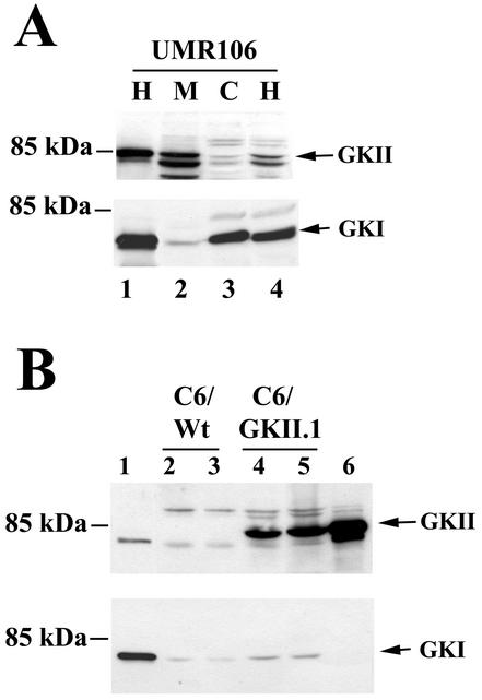
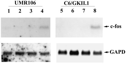
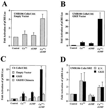
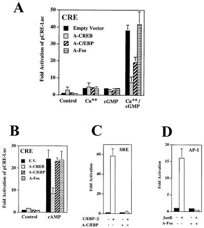
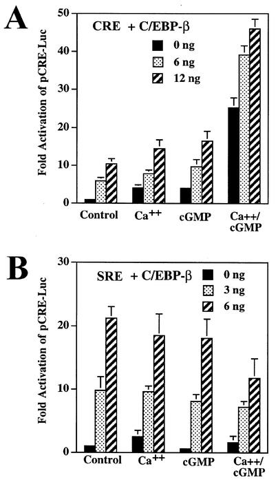
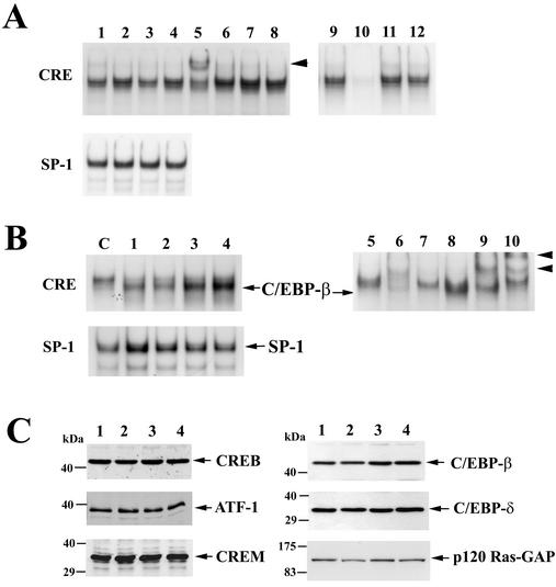
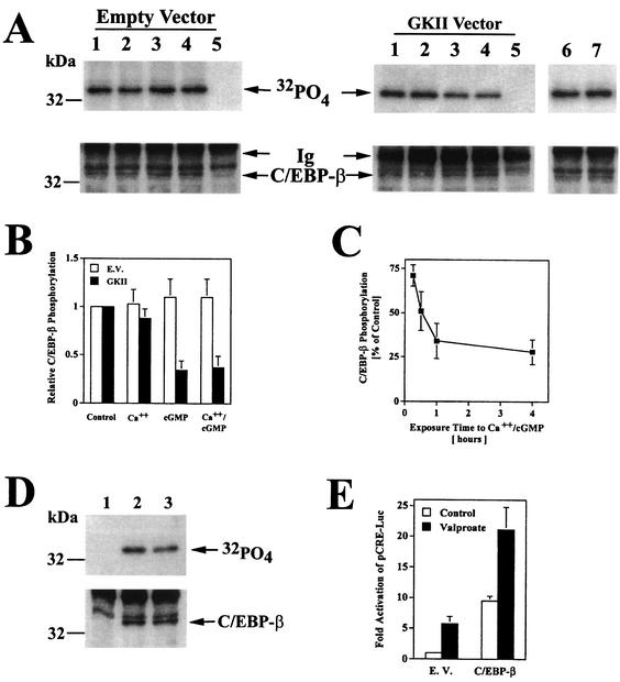
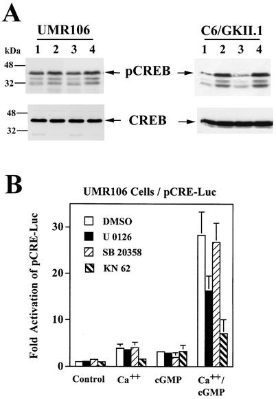
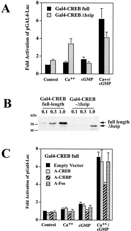
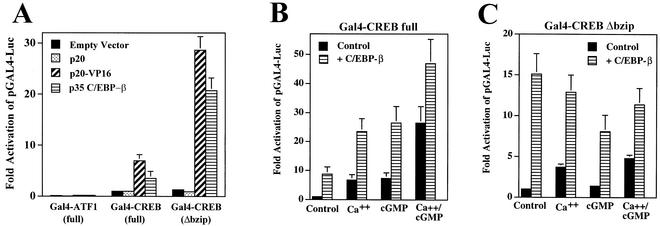
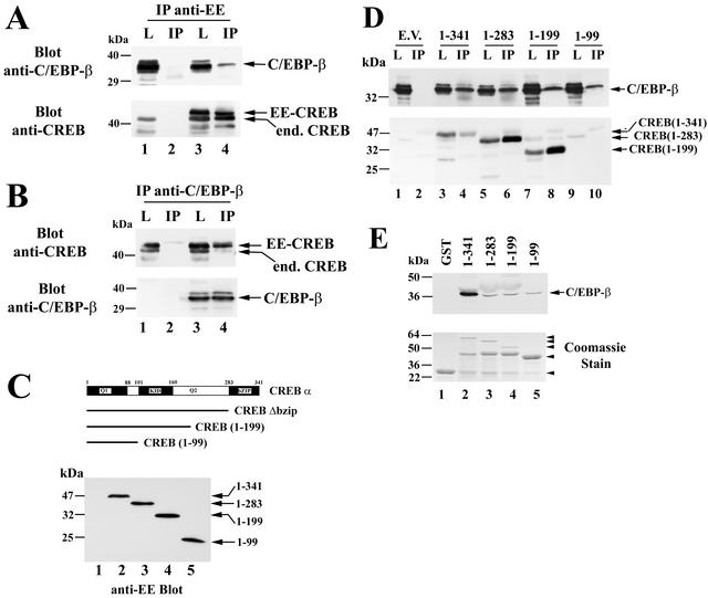
Calcium induces transcriptional activation of the fos promoter by activation of the cyclic AMP response element (CRE)-binding protein (CREB), and in some cells its effect is enhanced synergistically by cyclic GMP (cGMP) through an unknown mechanism. We observed calcium-cGMP synergism in neuronal and osteogenic cells which express type II cGMP-dependent protein kinase (G-kinase); the effect on the fos promoter was mediated by the CRE and proportional to G-kinase activity. Dominant negative transcription factors showed involvement of CREB- and C/EBP-related proteins but not of AP-1. Expression of C/EBP-beta but not C/EBP-alpha or -delta enhanced the effects of calcium and cGMP on a CRE-dependent reporter gene. The transactivation potential of full-length CREB fused to the DNA-binding domain of Gal4 was increased synergistically by calcium and cGMP, and overexpression of C/EBP-beta enhanced the effect, while a dominant negative C/EBP inhibited it. With a mammalian two-hybrid system, coimmunoprecipitation experiments, and in vitro binding studies, we demonstrated that C/EBP-beta and CREB interacted directly; this interaction involved the C terminus of C/EBP-beta but occurred independently of CREB's leucine zipper domain. CREB Ser(133) phosphorylation was stimulated by calcium but not by cGMP; in cGMP-treated cells, (32)PO(4) incorporation into C/EBP-beta was decreased and C/EBP-beta/CRE complexes were increased, suggesting regulation of C/EBP-beta functions by G-kinase-dependent dephosphorylation. C/EBP-beta and CREB associated with the fos promoter in intact cells, and the amount of promoter-associated C/EBP-beta was increased by calcium and cGMP. We conclude that calcium and cGMP transcriptional synergism requires cooperation of CREB and C/EBP-beta, with calcium and cGMP modulating the phosphorylation states of CREB and C/EBP-beta, respectively.


Figures

FIG. 1.
FIG. 1.
Expression of G-kinases I and II in UMR106 and C6 cells. (A) UMR106 cells were transfected with expression vectors encoding either G-kinase II (GKII, lane 1, upper panel) or G-kinase I (GKI, lane 1, lower panel) or left untransfected (lanes 2 to 4). Postnuclear homogenates (H), membranes (M), and cytosol (C) were prepared as described in Materials and Methods, and proteins were resolved by SDS-PAGE; Western blots were developed with antibodies specific for G-kinase II (upper panel) or G-kinase I (lower panel). The faster-migrating bands in the upper panel likely represent degradation products of G-kinase II, as they were decreased when denaturing SDS sample buffer was added immediately after cell lysis, as shown in lane 1. (B) C6 cells were transiently transfected with expression vectors encoding G-kinase I (lane 1) or G-kinase II (lane 6) or left untransfected (C6/Wt, lanes 2 and 3); C6 cells stably transfected with G-kinase II are shown for comparison (C6/GKII.1, lanes 4 and 5). Whole-cell homogenates were analyzed by Western blotting as described above.
FIG. 2.
FIG. 2.
Calcium and cGMP synergistically induce c-fos mRNA in UMR106 cells and G-kinase II-expressing C6 cells. Untransfected UMR106 cells (lanes 1 to 4) and stably transfected C6 cells expressing G-kinase II (C6/GKII.1, lanes 5 to 8) were cultured in DMEM containing 0.1% FBS and 0.1% BSA for 24 h; cells were treated for 30 min with buffer (lanes 1 and 5), calcium ionophore A23187 (0.3 μM, lanes 2 and 6), CPT-cGMP (250 μM, lanes 3 and 7), or both A23187 and CPT-cGMP (lanes 4 and 8). Northern blots were prepared with 30 μg (UMR106) or 20 μg (C6/GKII) of total cytoplasmic RNA per lane and hybridized to a c-fos cDNA probe as described in Materials and Methods (upper panels). Blots were reprobed with a cDNA probe encoding glyceraldehyde-3-phosphate dehydrogenase to demonstrate equal loading (GAPD, lower panels).
FIG. 3.
FIG. 3.
Synergistic activation of CRE- but not SRE-driven reporter genes by calcium and cGMP. UMR106 (A, B, and D) and C6 cells (C) were transiently transfected with the reporter plasmid pCRE-Luc (A to C) or pSRE-Luc (D) and with the control plasmid pRSV-βGal; cells were cotransfected with either empty vector (A, C, and D), G-kinase II expression vector (B to D), or a membrane-targeted G-kinase I construct (G-kinase I/II chimera; C) as described in Materials and Methods. Cells were maintained in low-serum medium for 24 h before they were treated for 8 h with 0.3 μM calcium ionophore A23187 (Ca++), 250 μM CPT-cGMP (cGMP), or both, as indicated. Luciferase and β-galactosidase activities were measured as described in Materials and Methods, and the ratio of luciferase to β-galactosidase activity of untreated cells was assigned a value of 1. Cotransfection of G-kinase II or the G-kinase I/II chimera had no significant effect in untreated cells.
FIG. 4.
FIG. 4.
Synergistic activation of CRE-dependent transcription by calcium and cGMP is prevented by dominant negative CREB and C/EBP constructs but not by a dominant negative Fos. (A and B) UMR106 cells were transfected with pCRE-Luc, pRSV-βGal, or G-kinase II vector as described in the legend to Fig. 3; some cells were cotransfected with 40 ng of expression vector encoding the dominant negative A-CREB, A-C/EBP, A-Fos, or empty vector. Cells were treated for 8 h with 0.3 μM A23187 (Ca++), 250 μM CPT-cGMP (cGMP), or both (A), or cells were treated with 100 μM CPT-cAMP (B), as indicated. The ratio of luciferase to β-galactosidase activity was normalized as described in the legend to Fig. 3. (C) Cells were transfected with pSRE-Luc, pRSV-βGal, and G-kinase II and cotransfected with empty vector (solid bars) or C/EBP-β (20 ng; open bars); some cells also received vector encoding A-C/EBP (40 ng) as indicated. (D) Cells were transfected with pAP1-Luc, pRSV-βGal, and G-kinase II and cotransfected with empty vector (solid bars) or JunB (20 ng; open bars); some cells also received vector encoding A-Fos (40 ng) as indicated.
FIG. 5.
FIG. 5.
C/EBP-β enhances the effects of calcium and cGMP on a CRE-dependent reporter. (A) UMR106 cells were transfected with pCRE-Luc, pRSV-βGal, and G-kinase II as described in the legend to Fig. 3; cells additionally received either empty vector (0 ng) or expression vector encoding C/EBP-β (6 ng and 12 ng). (B) Cells were transfected as in panel A except that pSRE-Luc was substituted for pCRE-luc and only 3 or 6 ng of C/EBP-β was cotransfected. Luciferase/β-galactosidase activity ratios were normalized as described in the legend to Fig. 3.
FIG. 6.
FIG. 6.
Effect of calcium and cGMP on CRE-binding proteins and expression of CREB- and C/EBP-related proteins. (A) UMR106 cells were left untreated (lanes 1 and 9 to 12) or treated for 4 h with 0.3 μM A23187 (lane 2), 250 μM CPT-cGMP (lane 3), or both drugs (lanes 4 to 8). Nuclear extracts were prepared, and electrophoretic mobility shift assays were performed with a radioactively labeled oligodeoxynucleotide probe encoding a CRE consensus sequence (upper panel) or an SP-1 consensus sequence (lower panel) as described in Materials and Methods. Some nuclear extracts were incubated with antibodies specific for CREB (lane 5), C/EBP-α (lane 6), C/EBP-β (lane 7), or control IgG (lane 8) prior to incubation with the CRE probe; the arrowhead indicates a supershifted complex obtained with anti-CREB antibody. To demonstrate competition of sequence-specific binding proteins, unlabeled CRE oligodeoxynucleotide (lane 10), SP-1 oligodeoxynucleotide (lane 11), or C/EBP consensus oligodeoxynucleotide (lane 12) was added at a 50-fold excess. (B) C6 cells were cotransfected with G-kinase II and either empty vector (lanes C and 5 to 7) or a vector encoding C/EBP-β (lanes 1 to 4 and 8 to 10); the cells in lanes 1 to 4 were treated as described for lanes 1 to 4 of panel A, and the cells in lane C and lanes 5 to 10 were treated with both A23187 and CPT-cGMP. Electrophoretic mobility shift assays were performed with the CRE probe (upper panel) or the SP-1 probe (lower panel) as described for panel A. Migration of a protein-DNA complex containing the transfected C/EBP-β is indicated in the upper panel. Some nuclear extracts were incubated with buffer (lanes 5 and 8) or with antibodies specific for CREB (lanes 6 and 9) or C/EBP-β (lanes 7 and 10) prior to incubation with the CRE probe. (C) UMR106 cells in lanes 1 to 4 were treated as described for lanes 1 to 4 of panel A. Equal amounts of nuclear extract protein were analyzed by SDS-PAGE and Western blotting with antibodies specific for CREB, ATF-1, CREM, C/EBP-β, C/EBP-δ, and p120 Ras-GTPase activating protein (GAP), serving as a control for contaminating cytoplasmic proteins.
FIG. 7.
FIG. 7.
Decreased C/EBP-β phosphorylation in cGMP-treated cells. (A) C6 cells were transfected with an expression vector encoding C/EBP-β and either empty vector (left panel, lanes 1 to 5), G-kinase II (right panel, lanes 1 to 5) or a G-kinase I/II chimera (right panel, lanes 6 and 7); cells were incubated with 32PO4 for 4 h and treated for 1 h with buffer (lanes 1, 5, and 6), 0.3 μM A23187 (lane 2), 250 μM CPT-cGMP (lane 3), or both agents (lanes 4 and 7) as described in Materials and Methods. Cell lysates were subjected to immunoprecipitation with a rabbit anti-C/EBP-β antibody (lanes 1 to 4, 6, and 7) or control rabbit IgG (lane 5). Immunoprecipitates were analyzed by SDS-PAGE, electroblotting, and autoradiography (upper panel) and by blotting with a murine anti-C/EBP-β antibody (lower panel). The immunoglobulin heavy chain band is labeled Ig. (B) 32PO4 incorporation into C/EBP-β was quantitated by scanning densitometry of autoradiographs of three independent experiments performed as described for panel A; cells were transfected with C/EBP-β plus either empty vector (open bars) or G-kinase II (solid bars). (C) Cells were transfected with C/EBP-β and G-kinase II and labeled for 4 h in 32PO4-containing medium as described for panel A. Half of the cultures were treated with 0.3 μM A23187 and 250 μM CPT-cGMP for the indicated times, and 32PO4 incorporation into C/EBP-β was quantitated as described for panel B. Data are expressed as percentages of the level in untreated controls. (D) Cells transfected with C/EBP-β were labeled with 32PO4 for 4 h and either left untreated (lanes 1 and 2) or treated for 1 h with 5 mM sodium valproate (lane 3); cell lysates were processed as described for panel A (lane 1, control IgG; lanes 2 and 3, anti-C/EBP-β antibody). (E) Cells were transfected with pCRE-Luc, pRSV-βGal, and either empty vector (E.V.) or 12 ng of C/EBP-β vector; cultures were either left untreated (open bars) or treated with 5 mM sodium valproate for 8 h (solid bars). Luciferase activity was normalized to β-galactosidase activity as described for Fig. 3.
FIG. 8.
FIG. 8.
Effect of calcium and cGMP on CREB phosphorylation and effect of Cam-kinase and MAP kinase inhibitors on calcium- and cGMP-stimulated transcription. (A) UMR106 cells (left panel) and C6/GKII.1 cells (right panel) were treated for 1 h with 0.3 μM A23187 (lane 2), 250 μM CPT-cGMP (lane 3), or both (lane 4). Equal amounts of cell extracts were analyzed by SDS-PAGE and Western blotting with an antibody specific for Ser133-phosphorylated CREB (pCREB, upper panel) and an antibody that recognizes CREB irrespective of its phosphorylation status (CREB, lower panel). The phospho-CREB antibody cross-reacts with phosphorylated ATF-1 and probably CREM (64). (B) UMR106 cells were transfected with pCRE-Luc, pRSV-βGal, and G-kinase II and treated with A23187 (Ca++), CPT-cGMP (cGMP), or both, as described in the legend to Fig. 3. At 24 h before harvesting, some cells were treated with 0.1% dimethyl sulfoxide (vehicle; DMSO), 10 μM U0126, 10 μM SB20358, or 10 μM KN62. Reporter gene activities were normalized as described for Fig. 3.
FIG. 9.
FIG. 9.
Calcium and cGMP synergistically increase the transactivation potential of full-length Gal4-CREB but not of Gal4-CREB-Δbzip; effect of A-C/EBP. (A) UMR106 cells were transfected with the reporter plasmid pGAL4-Luc, pRSV-βGal, and G-kinase II; cells were cotransfected with a vector encoding either full-length Gal4-CREB or Gal4-CREB-Δbzip. Cells were treated with A23187 (Ca++) and/or CPT-cGMP (cGMP), and reporter gene activities were measured as described in the legend to Fig. 3. The luciferase/β-galactosidase activity ratio of untreated cells transfected with full-length Gal4-CREB was assigned a value of 1. We transfected different amounts of vector encoding full-length Gal4-CREB (15 ng) or Gal4-CREB-Δbzip (5 ng) to produce similar reporter gene activities in untreated cells. When the same amount of each vector was transfected, the activity of Gal4-CREB-Δbzip was 2.8- ± 0.4-fold higher than the activity of full-length Gal4-CREB (not shown). (B) In parallel experiments, cells were transfected with 0.1 μg, 0.3 μg, or 1 μg of full-length Gal4-CREB or Gal4-CREB-Δbzip, as indicated, to examine the expression levels of both constructs by Western blotting with an antibody specific for the Gal4 DNA-binding domain. (C) UMR106 cells were transfected with full-length Gal4-CREB, pGAL4-Luc, pRSV-βGal, and G-kinase II as described for panel A; cells were cotransfected with either empty vector or 40 ng of expression vector encoding A-CREB, A-C/EBP, or A-Fos. Cells were treated, and luciferase/β-galactosidase activity ratios were determined as described for panel A.
FIG. 10.
FIG. 10.
C/EBP-β enhances the transactivation potential of Gal4-CREB and Gal4-CREB-Δbzip. (A) UMR106 cells were transfected with pGAL4-Luc, pRSV-βGal, and Gal4-ATF-1 (100 ng), Gal4-CREB (15 ng), or Gal4-CREB-Δbzip (5 ng) as indicated; cells were cotransfected with either empty vector, 100 ng of expression vector encoding p20, 100 ng of p20 fused to the activation domain of VP16, or 12 ng of full-length C/EBP-β (p35). Different amounts of vector encoding p20, p20-VP16, and p35 were used because Western blotting with an antibody specific for the C terminus of C/EBP-β showed that the p20 and p20-VP16 vectors expressed about eightfold-lower protein levels than the same amount of p35 C/EBP-β vector (not shown). The luciferase/β-galactosidase activity ratio of untreated cells transfected with full-length Gal4-CREB was assigned a value of 1. (B and C) C6 cells were transfected with pGAL4-Luc, pRSV-βGal, G-kinase II, and either full-length Gal4-CREB (15 ng, panel B) or pGal4-CREBΔbzip (5 ng, panel C); cells were cotransfected with either empty vector (control) or 12 ng of expression vector encoding C/EBP-β. Cells were treated with A23187 (Ca++) and/or CPT-cGMP (cGMP), and luciferase/β-galactosidase activity ratios were determined as described in the legend to Fig. 3.
FIG. 11.
FIG. 11.
CREB and C/EBP-β association in vitro and in intact cells. (A) COS7 cells were electroporated with C/EBP-β vector (lanes 1 to 4) and either empty vector (lanes 1 and 2) or vector encoding EE epitope-tagged CREB (lanes 3 and 4). Cell lysates were subjected to immunoprecipitation with an anti-EE epitope antibody and washed immunoprecipitates (IP) or 5% of the input lysates (L) were analyzed by SDS-PAGE and Western blotting. Blots were developed with antibodies specific for C/EBP-β (upper panel) and reprobed with anti-CREB antibody (lower panel). Note the presence of endogenous CREB in cell lysates (lanes 1 and 3) as well as in immunoprecipitates containing EE-CREB, which heterodimerizes with endogenous CREB (lane 4); the lowest band most likely represents a proteolytic breakdown product, as it varied in intensity in different experiments. (B) COS7 cells were electroporated with EE-CREB (lanes 1 to 4) and either empty vector (lanes 1and 2) or vector encoding C/EBP-β (lanes 3 and 4). Immunoprecipitates (IP) were obtained with a C/EBP-β-specific antibody and analyzed side by side with 5% of input lysates (L) by Western blotting with antibodies specific for CREB (upper panel) and C/EBP-β (lower panel). Note that endogenous C/EBP-β in COS cells migrates with an apparent molecular mass of 42 to 45 kDa and is only visualized on longer exposures (not shown). (C) COS7 cells were cotransfected with C/EBP-β and either empty vector (lane 1) or vectors encoding EE epitope-tagged full-length CREB (lane 2), CREBΔbzip (lane 3), CREB truncated at amino acid 199 (lane 4), or CREB truncated at amino acid 99 (lane 5). Western blots were developed with anti-EE antibody. (D) Cell lysates of the COS7 cells described for panel C were subjected to immunoprecipitation with anti-EE antibody as described for panel A, and the immunoprecipitates (IP) as well as 5% of cell lysate input (L) were analyzed by Western blotting with an anti-C/EBP-β-specific antibody (upper panel) or a CREB-specific antibody (lower panel). Note that the anti-CREB antibody does not recognize the construct encoding amino acids 1 to 99 (lanes 9 and 10), but expression of the protein was demonstrated on the anti-EE blot shown in panel C. (E) GST (lane 1) or GST fusion proteins encoding full-length CREB or the indicated C-terminal CREB truncations (lanes 2 to 5) were bound to glutathione-agarose beads and incubated with purified Myc epitope-tagged C/EBP-β as described in Materials and Methods. Washed beads were analyzed by Western blotting with anti-Myc antibody to detect C/EBP-β (upper panel). An aliquot of the GST proteins was analyzed by SDS-PAGE and Coomassie staining (lower panel). The arrowheads indicate the migration of full-length proteins; in lanes 2 to 4, an ≈45-kDa proteolytic breakdown product is present.
FIG. 12.
FIG. 12.
Association of CREB and C/EBP-β with the fos promoter in intact cells. (A) UMR106 cells were cultured for 4 h in the absence (lanes 1, 3, and 5 to 7) or presence (lanes 2 and 4) of 0.3 μM A23187 and 250 μM CPT-cGMP. After a brief treatment with 1% formaldehyde to cross-link proteins and DNA, cells were lysed, and chromatin was sheared as described in Materials and Methods. These cell lysates were subjected to immunoprecipitation with antibodies specific for CREB (lanes 1 and 2), C/EBP-β (lanes 3 and 4), or control IgG (lane 5). DNA was isolated from the immunoprecipitates and amplified with radioactively labeled primers flanking the CRE of the fos promoter; amplification products were analyzed by nondenaturing SDS-PAGE and autoradiography. Lanes 6 and 7 show amplification products obtained with a fraction of the cell lysate, corresponding to 10−5 (lane 6) and 3 × 10⁻⁶ (lane 7) of the input. (B) Densitometric analysis of autoradiographs from three independent experiments performed as described for panel A.

Similar articles

Cited by

References

    1. Aguirre, J., L. Buttery, M. O'Shaughnessy, F. Afzal, I. F. de Marticorena, M. Hukkanen, P. Huang, I. Maclntyre, and J. Polak. 2001. Endothelial nitric oxide synthase gene-deficient mice demonstrate marked retardation in postnatal bone formation, reduced bone volume, and defects in osteoblast maturation and activity. Am. J. Pathol. 158:247-257. - PMC - PubMed
    1. Ahn, S., M. Olive, S. Aggarwal, D. Krylov, D. D. Ginty, and C. Vinson. 1998. A dominant-negative inhibitor of CREB reveals that it is a general mediator of stimulus-dependent transcription of c-fos. Mol. Cell. Biol. 18:967-977. - PMC - PubMed
    1. Arancio, O., I. Antonova, S. Gambaryan, S. M. Lohmann, J. S. Wood, D. S. Lawrence, and R. D. Hawkins. 2001. Presynaptic role of cGMP-dependent protein kinase during long-lasting potentiation. J. Neurosci. 21:143-149. - PMC - PubMed
    1. Belsham, D. D., and P. L. Mellon. 2000. Transcription factors Oct-1 and C/EBPβ (CCAAT/enhancer-binding protein β) are involved in the glutamate/nitric oxide/cyclic-guanosine 5′-monophosphate-mediated repression of gonadotropin-releasing hormone gene expression. Mol. Endocrinol. 14:212-228. - PubMed
    1. Belsham, D. D., W. C. Wetsel, and P. L. Mellon. 1996. NMDA and nitric oxide act through the cGMP signal transduction pathway to repress hypothalamic gonadotropin-releasing hormone gene expression. EMBO J. 15:538-547. - PMC - PubMed

Publication types

MeSH terms

LinkOut - more resources