. 2003 Jun;23(12):4207-18.
doi: 10.1128/MCB.23.12.4207-4218.2003.

Methylation of histone H3 by Set2 in Saccharomyces cerevisiae is linked to transcriptional elongation by RNA polymerase II

Affiliations

Methylation of histone H3 by Set2 in Saccharomyces cerevisiae is linked to transcriptional elongation by RNA polymerase II

Nevan J Krogan et al. Mol Cell Biol. 2003 Jun.

Abstract

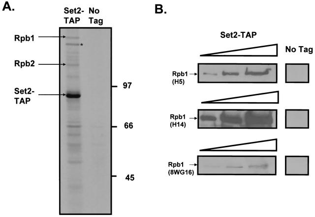
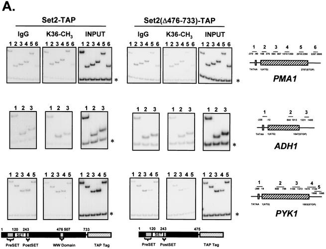
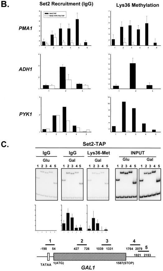
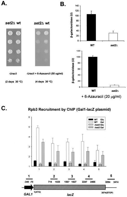
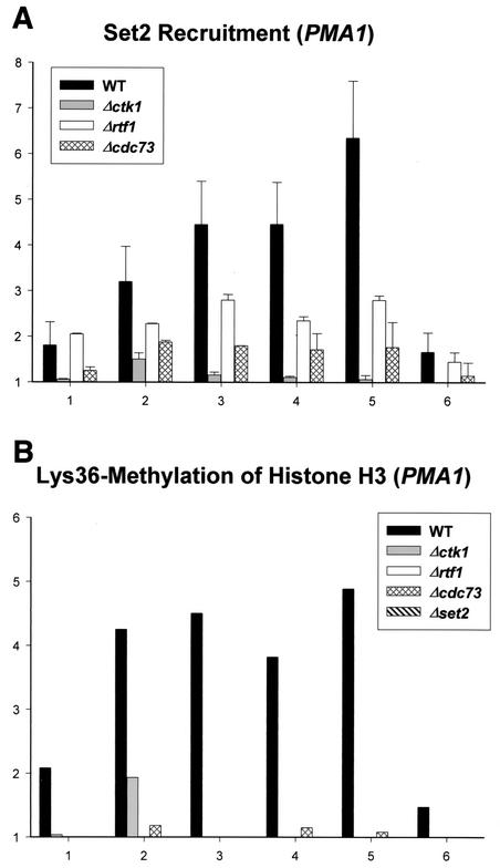
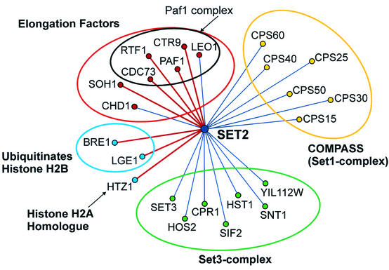
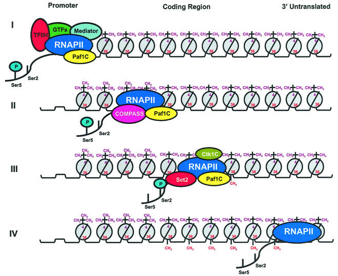
Set2 methylates Lys36 of histone H3. We show here that yeast Set2 copurifies with RNA polymerase II (RNAPII). Chromatin immunoprecipitation analyses demonstrated that Set2 and histone H3 Lys36 methylation are associated with the coding regions of several genes that were tested and correlate with active transcription. Both depend, as well, on the Paf1 elongation factor complex. The C terminus of Set2, which contains a WW domain, is also required for effective Lys36 methylation. Deletion of CTK1, encoding an RNAPII CTD kinase, prevents Lys36 methylation and Set2 recruitment, suggesting that methylation may be triggered by contact of the WW domain or C terminus of Set2 with Ser2-phosphorylated CTD. A set2 deletion results in slight sensitivity to 6-azauracil and much less beta-galactosidase produced by a reporter plasmid, resulting from a defect in transcription. In synthetic genetic array (SGA) analysis, synthetic growth defects were obtained when a set2 deletion was combined with deletions of all five components of the Paf1 complex, the chromodomain elongation factor Chd1, the putative elongation factor Soh1, the Bre1 or Lge1 components of the histone H2B ubiquitination complex, or the histone H2A variant Htz1. SET2 also interacts genetically with components of the Set1 and Set3 complexes, suggesting that Set1, Set2, and Set3 similarly affect transcription by RNAPII.


Figures

FIG. 1.
FIG. 1.
TAP of Set2. (A) Purification of Set2 was carried out with strains containing either no tagged protein or a TAP-tagged version of Set2. The protein complex was purified in the presence of 100 mM NaCl as described in the text and was then analyzed by SDS-PAGE and silver staining. Set2 and subunits of RNAPII were identified by trypsin digestion and MALDI-TOF. The asterisk indicates another polypeptide, Rrp5, which was also identified in this preparation and is most likely a contaminant. (B) Immunoprecipitation with IgG followed by Western blot analysis using the monoclonal antibodies H5 and H14 (6, 40) demonstrated that Set2 copurified with RNAPII phosphorylated on both Ser2 and Ser5 in the CTD repeats of Rpb1. The RNAPII that copurified with Set2 also reacted with the antibody 8WG16 (60), which recognizes both partially phosphorylated and unphosphorylated Rpb1. Western blotting analyses with immunoprecipitates using extracts from an untagged strain were also performed as controls with all three antibodies. Successive lanes were loaded with 0.5, 1.5, and 2.5 μg of extract protein.
FIG. 2.
FIG. 2.
Set 2 and histone H3 methylation on Lys36 localize to transcribed regions. (A) ChIP was performed to monitor the presence of either the Set2 protein or Lys36 methylation on histone H3 along the PMA1, ADH1, and PYK1 genes. Chromatin was immunoprecipitated either with rabbit IgG-agarose from strains containing TAP-tagged versions of Set2 or with antibody against Lys36 methylated histone H3 (Upstate Biotechnology) from a strain with no tag. The Set2(Δ476-733)-TAP strain was constructed by recombining in the TAP tag so as to remove the last 258 amino acids of Set2. PCR amplification was carried out by using primer pairs recognizing promoter (lane 1), coding (lanes 2, 3, 4, and 5), and 3′ untranslated (lane 6) regions for PMA1; promoter (lane 1), coding (lane 2), and 3′ untranslated (lane 3) regions for ADH1; and promoter (lane 1), coding (lanes 2, 3, and 4), and 3′ untranslated regions for PYK1. Each PCR contained a second primer pair that amplified a region of chromosome V devoid of open reading frames (marked by asterisks), thus providing an internal control for background. Input, signal from chromatin before immunoprecipitation. Primer pairs used are as follows: for PMA1, PMA1−370 and PMA1−47 (lanes 1), PMA1168 and PMA1376 (lanes 2), PMA1584 and PMA1807 (lane 3), PMA11010 and PMA11250 (lanes 4), PMA12018 and PMA12290 (lanes 5), and PMA13287 and PMA13500 (lanes 6); for ADH1, ADH1−235 and ADH1−13 (lanes 1), ADH1844 and ADH11013 (lanes 2), and ADH11231 and ADH11400 (lanes 3); for PYK1, PYK1−288 and PYK119 (lanes 1), PYK1508 and PYK1799 (lanes 2), PYK11104 and PYK11372 (lanes 3), PYK11470 and PYK11720 (lanes 4), and PYK11695 and PYK11995 (lanes 5); for the nontranscribed region, Intergenic V-1 and Intergenic V-2. (B) Quantitation of the ChIP assays. Each value is calculated by dividing the ratio of the ChIP signal to the input signal for the experimental PCR product by the ratio of the ChIP signal to the input signal for the control PCR product. (C) Set2 is recruited to active genes. Cells grown overnight in medium containing 2% glucose were harvested, washed twice with sterile water, and inoculated at an OD595 of ≈0.02 into 300 ml of medium containing either 2% glucose or 2% galactose and 1% raffinose. Cells were incubated at 30°C until the OD595 reached 0.6 and treated with 1% formaldehyde for ChIP assays. Primer pairs used for PCR are as follows: GAL1−190 and GAL154 (lanes 1), GAL1427 and GAL1726 (lanes 2), GAL11039 and GAL11331 (lanes 3), GAL11764 and GAL12079 (lanes 4), and GAL11921 and GAL12153 (lanes 5); for the nontranscribed region, Intergenic V-1 and Intergenic V-2.
FIG. 2.
FIG. 2.
Set 2 and histone H3 methylation on Lys36 localize to transcribed regions. (A) ChIP was performed to monitor the presence of either the Set2 protein or Lys36 methylation on histone H3 along the PMA1, ADH1, and PYK1 genes. Chromatin was immunoprecipitated either with rabbit IgG-agarose from strains containing TAP-tagged versions of Set2 or with antibody against Lys36 methylated histone H3 (Upstate Biotechnology) from a strain with no tag. The Set2(Δ476-733)-TAP strain was constructed by recombining in the TAP tag so as to remove the last 258 amino acids of Set2. PCR amplification was carried out by using primer pairs recognizing promoter (lane 1), coding (lanes 2, 3, 4, and 5), and 3′ untranslated (lane 6) regions for PMA1; promoter (lane 1), coding (lane 2), and 3′ untranslated (lane 3) regions for ADH1; and promoter (lane 1), coding (lanes 2, 3, and 4), and 3′ untranslated regions for PYK1. Each PCR contained a second primer pair that amplified a region of chromosome V devoid of open reading frames (marked by asterisks), thus providing an internal control for background. Input, signal from chromatin before immunoprecipitation. Primer pairs used are as follows: for PMA1, PMA1−370 and PMA1−47 (lanes 1), PMA1168 and PMA1376 (lanes 2), PMA1584 and PMA1807 (lane 3), PMA11010 and PMA11250 (lanes 4), PMA12018 and PMA12290 (lanes 5), and PMA13287 and PMA13500 (lanes 6); for ADH1, ADH1−235 and ADH1−13 (lanes 1), ADH1844 and ADH11013 (lanes 2), and ADH11231 and ADH11400 (lanes 3); for PYK1, PYK1−288 and PYK119 (lanes 1), PYK1508 and PYK1799 (lanes 2), PYK11104 and PYK11372 (lanes 3), PYK11470 and PYK11720 (lanes 4), and PYK11695 and PYK11995 (lanes 5); for the nontranscribed region, Intergenic V-1 and Intergenic V-2. (B) Quantitation of the ChIP assays. Each value is calculated by dividing the ratio of the ChIP signal to the input signal for the experimental PCR product by the ratio of the ChIP signal to the input signal for the control PCR product. (C) Set2 is recruited to active genes. Cells grown overnight in medium containing 2% glucose were harvested, washed twice with sterile water, and inoculated at an OD595 of ≈0.02 into 300 ml of medium containing either 2% glucose or 2% galactose and 1% raffinose. Cells were incubated at 30°C until the OD595 reached 0.6 and treated with 1% formaldehyde for ChIP assays. Primer pairs used for PCR are as follows: GAL1−190 and GAL154 (lanes 1), GAL1427 and GAL1726 (lanes 2), GAL11039 and GAL11331 (lanes 3), GAL11764 and GAL12079 (lanes 4), and GAL11921 and GAL12153 (lanes 5); for the nontranscribed region, Intergenic V-1 and Intergenic V-2.
FIG. 3.
FIG. 3.
Set2 influences transcription by RNAPII. (A) Sensitivity of the set2 deletion strain to 6-AU. Strains containing either wild-type SET2 or a set2Δ allele, as well as the plasmid pRS316 (51), were plated on synthetic dextrose-uracil medium with or without 6-AU (50 μg/ml) and were grown at 30°C for 2 to 4 days. WT, wild type. (B) Expression of β-galactosidase from the lacZ fusion plasmid p416GAL1-lacZ (9) in WT and set2Δ cells. Cells were grown in medium containing 2% glucose until an OD595 of ≈1 was reached and washed three times in distilled water; then, medium containing 2% galactose was added with or without 20 μg of 6-AU/ml and β-galactosidase activities were assayed after 4 h of growth at 30°C. (C) Density of RNAPII along the lacZ gene on the GAL1-lacZ plasmid. By employing an antibody that recognizes the Rpb3 subunit of RNAPII (Neoclone Biotechnology), ChIP was used to monitor the approximate relative concentrations of RNAPII at various positions across the lacZ gene. Wild-type and set2Δ strains harboring p416GAL1-lacZ were incubated in medium containing 2% glucose until an OD595 of 1 was reached. Cells were harvested, washed three times with water, and reinoculated into media containing either 2% glucose or 2% galactose. After 4 h of incubation at 30°C, cells were cross-linked with formaldehyde for the ChIP assays. PCR amplification was carried out with primer pairs recognizing promoter (labeled 1), coding (labeled 2, 3, and 4) and 3′ untranslated (labeled 5) regions for GAL1-lacZ. Primer pairs used for PCR were as follows: 1, GAL1-lacZ-340 and GAL-lacZ−70; 2, GAL1-lacZ715 and GAL1-lacZ1025; 3, GAL1-lacZ1567 and GAL1-lacZ1827; 4, GAL1-lacZ2247 and GAL1-lacZ2465; and 5, GAL1-lacZ3094 and GAL1-lacZ3414; for the nontranscribed region, Intergenic V-1 and Intergenic V-2.
FIG. 4.
FIG. 4.
Effects of deleting genes encoding elongation factors on Set2 recruitment and histone H3 Lys36 methylation. (A) Recruitment of Set2 to an actively transcribed gene in strains containing deletions of genes encoding elongation factors. ChIP was used in strains containing TAP tags on Set2 to monitor the presence of Set2 along the PMA1 gene. Recruitment of Set2 is severely compromised when genes encoding subunits of the Paf1 complex, Rtf1 and Cdc73, or the RNAPII CTD kinase, Ctk1, are deleted. (B) The presence of Lys36 methylation on histone H3 in strains containing deletions of genes encoding elongation factors. Deletions of CDC73, RTF1, and CTK1, as well as SET2, eliminate Lys36 methylation on the PMA1 gene.
FIG. 5.
FIG. 5.
Genetic interaction network representing the synthetic growth defects identified by SGA analysis. Genes are represented by nodes, and interactions are represented by lines that connect the nodes. All of the interactions were confirmed by either tetrad analysis (for synthetic lethals) or random sporulation (for synthetic growth defects). Synthetic lethal interaction are shown as red lines, and synthetic growth defects are shown as blue lines. There were approximately 40 other genetic interactions with SET2 that were identified in the screen and are not shown in this diagram.
FIG. 6.
FIG. 6.
Model for the coupling of histone methylation to transcriptional elongation and the phosphorylation of RNAPII in S. cerevisiae. See the text for details.

Similar articles

Cited by

References

    1. Allen, C., N. A. Kent, H. S. Jones, J. O'Sullivan, A. Aranda, and N. J. Proudfoot. 2002. A role for chromatin remodeling in transcriptional termination by RNA polymerase II. Mol. Cell 10:1442-1452. - PubMed
    1. Allison, L. A., M. Moyle, M. Shales, and C. J. Ingles. 1985. Extensive homology among the largest subunits of eukaryotic and prokaryotic RNA polymerases. Cell 42:599-610. - PubMed
    1. Archambault, J., F. Lacroute, A. Ruet, and J. D. Friesen. 1992. Genetic interaction between transcription elongation factor TFIIS and RNA polymerase II. Mol. Cell. Biol. 12:4142-4152. - PMC - PubMed
    1. Bähler, J., J. Wu, M. S. Longtine, N. G. Shah, A. McKenzie III, A. B. Steever, A. Wach, P. Philippsen, and J. R. Pringle. 1998. Heterologous modules for efficient and versatile PCR-based gene targeting in Schizosaccharomyces pombe. Yeast 14:943-951. - PubMed
    1. Bernstein, B. E., E. L. Humphrey, R. L. Erlich, R. Schneider, P. Bouman, J. S. Liu, T. Kouzarides, and S. L. Schreiber. 2002. Methylation of histone H3 Lys4 in coding regions of active genes. Proc. Natl. Acad. Sci. USA 99:8695-8700. - PMC - PubMed

Publication types

MeSH terms