. 2003 Jun;23(12):4257-66.
doi: 10.1128/MCB.23.12.4257-4266.2003.

Cysteine 64 of Ref-1 is not essential for redox regulation of AP-1 DNA binding

Affiliations

Cysteine 64 of Ref-1 is not essential for redox regulation of AP-1 DNA binding

Jared M Ordway et al. Mol Cell Biol. 2003 Jun.

Abstract

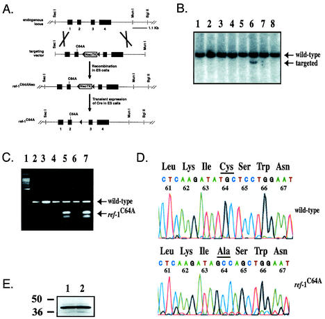
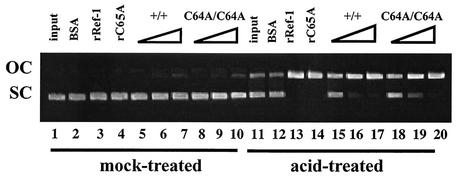
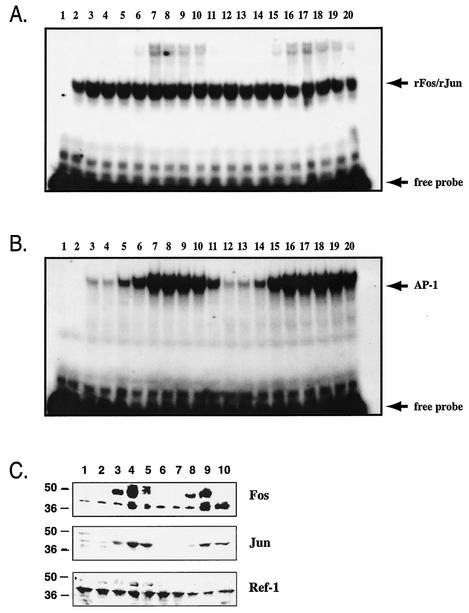
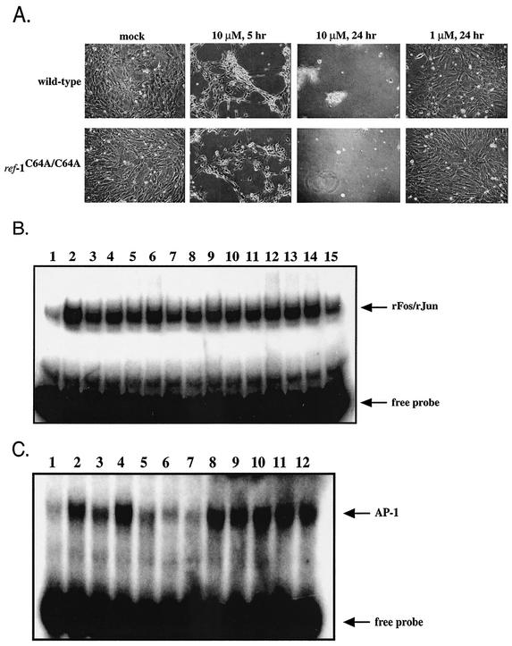
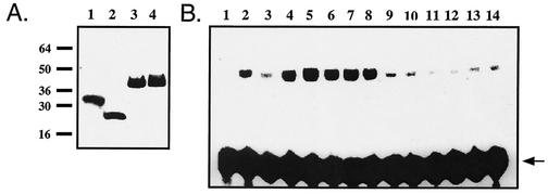
Ref-1 participates in DNA repair as well as in redox regulation of transcription factor function. The redox function of Ref-1 involves reduction of oxidized cysteine residues within the DNA binding domains of several transcription factors, including Fos and Jun. Reduction of these residues is required for DNA binding, providing a redox-dependent mechanism for regulation of target gene expression. Previous in vitro studies implicated cysteine 65 of human Ref-1 (cysteine 64 of mouse Ref-1) as the redox catalytic site. We analyzed the in vivo role of cysteine 64 in redox regulation of AP-1 activity by introducing a cysteine-to-alanine point mutation into the endogenous mouse Ref-1 gene (ref-1(C64A)). Unlike Ref-1 null mice, which die very early in embryonic development, homozygous ref-1(C64A) mice are viable, they survive to normal life expectancy, and they display no overt abnormal phenotype. Although Ref-1 provides the major AP-1-reducing activity in murine cells, ref-1(C64A) cells retain normal levels of endogenous AP-1 DNA binding activity in vivo as well as normal Fos- and Jun-reducing activity in vitro. These results demonstrate that Ref-1 cysteine 64/65 is not required for redox regulation of AP-1 DNA binding in vivo, and they challenge previous hypotheses regarding the mechanism by which Ref-1 regulates the redox-dependent activity of specific transcription factors.


Figures

FIG. 1.
FIG. 1.
(A) Diagram of gene-targeted mutagenesis strategy. See the text for details. ref-1C64ANeo, ref-1C64A with neomycin resistance cassette. (B) Southern blot screen for gene-targeted ES cell clones. DNA from neomycin-resistant ES cell clones was digested with SacI and BglII and hybridized to an external probe as shown in panel A. The NeoTK cassette includes a SacI site, allowing detection of homologous recombination by the presence of a 9.1-kb fragment in addition to the wild-type 10.8-kb fragment. Lane 6 represents a gene-targeted clone. (C) Confirmation of gene targeting by PCR followed by restriction enzyme digestion. Exon 2 was PCR amplified from ES cell DNA, and amplicons were digested with PvuII. Lanes: 1, DNA ladder; 2, wild-type undigested amplicons; 3, wild-type PvuII-digested amplicons; 4 and 6, ref-1C64A/+ undigested amplicons; 5 and 7, ref-1C64A/+ PvuII-digested amplicons. Arrows mark bands amplified from wild-type alleles and digested bands amplified from ref-1C64A alleles as indicated. (D) Confirmation of the endogenous ref-1C64A mutation by genomic sequencing. Ref-1 exon 2 was PCR amplified from genomic DNA extracted from wild-type and ref-1C64A/C64A primary embryonic fibroblasts, and amplicons were sequenced. Codons are labeled by mouse Ref-1 amino acid number and identity. Cysteine/alanine 64 is underlined. Sequencing verified the TG-GC mutation at codon 64 and the silent TC-AG mutation at codon 65. (E) Western blot analysis of Ref-1 protein in wild-type and ref-1C64A/C64A cells. Lanes: 1, 20 μg of wild-type embryonic fibroblast nuclear extract; 2, 20 μg of ref-1C64A/C64A embryonic fibroblast nuclear extract. Western blot analysis was performed with a polyclonal antibody specific for Ref-1 (43).
FIG. 2.
FIG. 2.
The C64A mutation does not affect Ref-1 AP endonuclease activity. Supercoiled pBluescript II plasmid was mock-treated or acid-treated to induce AP lesions as indicated. Plasmids were reacted with 50 ng of bovine serum albumin (BSA; lanes 2 and 12), 50 ng of recombinant human Ref-1 (rRef-1; lanes 3 and 13), 50 ng of recombinant human C65A Ref-1 (rC65A; lanes 4 and 14), nuclear extract from wild-type primary embryonic fibroblasts (10, 50, and 100 ng [lanes 5 to 7 and 15 to 17, respectively]), or nuclear extract from ref-1C64A/C64A primary embryonic fibroblasts (10, 50, and 100 ng [lanes 8 to 10 and 18 to 20, respectively]). Ref-1 AP endonuclease activity was assayed by the conversion of supercoiled plasmid (SC) to an open circular form (OC).
FIG. 3.
FIG. 3.
Ref-1C64A/C64A tissues retain normal Fos/Jun-reducing activity. Oxidized recombinant Fos and Jun peptides were incubated to allow heterodimerization. Extracts were added and Fos/Jun DNA binding was determined by EMSA by using a radiolabeled oligonucleotide probe containing an AP-1 DNA binding site. Lanes: 1, dilution buffer alone; 2, 10 mM DTT; 3 to 5, 10, 25, and 50 ng of wild-type lung extract, respectively; 6 to 8, 10, 25, and 50 ng of ref-1C64A/C64A lung extract, respectively; 9 to 11, 10, 25, and 50 ng of wild-type heart extract, respectively; 12 to 14, 10, 25, and 50 ng of ref-1C64A/C64A heart extract, respectively. Free probe is indicated by an arrow.
FIG. 4.
FIG. 4.
(A) Serum induction does not affect Fos/Jun-reducing activity in wild-type or ref-1C64A/C64A primary embryonic fibroblast extracts. Fos/Jun-reducing activity was determined by EMSA as described in the legend to Fig. 3. Primary embryonic fibroblasts were cultured in medium containing 0.5% FCS for 4 days and then stimulated with medium containing 20% FCS. Cells were harvested at various time points throughout the 24-h stimulation. Lanes: 1, dilution buffer alone; 2, 10 mM DTT; 3 to 11, 1-μg wild-type nuclear extracts harvested at 0, 0.2, 0.5, 1, 2, 3, 5, 10, and 24 h poststimulation, respectively; 12 to 20, 1-μg ref-1C64A/C64A nuclear extracts harvested at 0, 0.2, 0.5, 1, 2, 3, 5, 10, and 24 h poststimulation, respectively. Free probe and recombinant Fos/Jun-bound probe (rFos/rJun) are indicated by arrows. (B) Serum-induced endogenous AP-1 DNA binding is not compromised in ref-1C64A/C64A cells. Nuclear extracts were analyzed by EMSA as described for panel A except without added recombinant Fos and Jun peptides. Lanes are identical to those described for panel A. Free probe and recombinant Fos/Jun- or endogenous AP-1-bound probe are indicated by arrows. (C) Fos, Jun, and Ref-1 protein levels at selected time points during serum induction. Extracts described for panels A and B were subjected to Western analysis with antibodies for the indicated proteins. Lanes: 1 to 5, wild-type extracts at 0, 0.5, 1, 3, and 5 h poststimulation, respectively; 6 to 10, ref-1C64A/C64A extracts at 0, 0.5, 1, 3, and 5 h poststimulation, respectively. Antibodies are indicated on the right of the panel. Molecular masses in kilodaltons are indicated on the left on the panel.
FIG. 5.
FIG. 5.
(A) Wild-type and ref-1C64A/C64A embryonic fibroblasts are equally susceptible to H2O2 treatment. Primary embryonic fibroblast cultures were treated with H2O2 at concentrations ranging from 100 nM to 1 mM for 24 h. Mock-treated cultures and cultures treated with 1 μM and 10 μM H2O2 are shown at 5 and 24 h posttreatment, as indicated. (B) H2O2 treatment does not affect Fos/Jun-reducing activity in wild-type or ref-1C64A/C64A primary embryonic fibroblast extracts. Recombinant Fos/Jun-reducing activity was determined by EMSA as described in the legend to Fig. 3. Lanes: 1, mock-treated sample; 2, 10 mM DTT; 3 to 8, wild-type embryonic fibroblast extracts at 0, 0.5, 1, 3, 6, and 24 h following 1 μM H2O2 challenge, respectively; 9 to 14, ref-1C64A/C64A embryonic fibroblast extracts at 0, 0.5, 1, 3, 6, and 24 h following 1 μM H2O2 challenge, respectively; 15, extract dilution buffer alone. The recombinant Fos/Jun complex (rFos/rJun) and free probe are indicated by arrows. (C) H2O2-induced AP-1 DNA binding is enhanced in ref-1C64A/C64A cells. Endogenous AP-1 DNA binding activity was determined as described in the legend to Fig. 3. Lanes: 1 to 6, wild-type embryonic fibroblasts assayed at 0, 0.5, 1, 3, 6, and 24 h following challenge with 1 μM H2O2; 7 to 12, ref-1C64A/C64A embryonic fibroblasts assayed at 0, 0.5, 1, 3, 6, and 24 h following challenge with 1 μM H2O2. Free probe and endogenous AP-1-bound probe are indicated by arrows.
FIG. 6.
FIG. 6.
(A) All recombinant proteins were expressed as six-His-tagged fusion proteins and purified as described in Materials and Methods. Ten micrograms of each recombinant protein was run on a sodium dodecyl sulfate-polyacrylamide gel and stained with Coomassie blue. Lanes: 1, wbFos (1); 2, TK550 Jun (22); 3, wild-type Ref-1; 4, C65A Ref-1. (B) Recombinant C65A Ref-1 retains Fos/Jun-reducing activity. Recombinant Ref-1 proteins were assayed for redox activity by EMSA as described in the legend to Fig. 3. Recombinant protein stocks were adjusted to 10 mg/ml to ensure equal carryovers of storage buffer. Lanes: 1, dilution buffer alone; 2, 10 mM DTT; 3 to 5, 10, 25, and 50 ng of recombinant Ref-1, respectively; 6 to 8, 10, 25, and 50 ng of recombinant C65A Ref-1, respectively; 9 to 11, 10, 25, and 50 ng of recombinant mDab-1, respectively; 12 to 14, storage buffer diluted 1:1,000, 1:400, and 1:200, respectively. Free probe is indicated by an arrow.

Similar articles

Cited by

References

    1. Abate, C., D. Luk, R. Gentz, F. J. Rauscher III, and T. Curran. 1990. Expression and purification of the leucine zipper and DNA-binding domains of Fos and Jun: both Fos and Jun contact DNA directly. Proc. Natl. Acad. Sci. USA 87:1032-1036. - PMC - PubMed
    1. Abate, C., L. Patel, F. J. Rauscher III, and T. Curran. 1990. Redox regulation of fos and jun DNA-binding activity in vitro. Science 249:1157-1161. - PubMed
    1. Beiqing, L., M. Chen, and R. L. Whisler. 1996. Sublethal levels of oxidative stress stimulate transcriptional activation of c-jun and suppress IL-2 promoter activation in Jurkat T cells. J. Immunol. 157:160-169. - PubMed
    1. Demple, B., T. Herman, and D. S. Chen. 1991. Cloning and expression of APE, the cDNA encoding the major human apurinic endonuclease: definition of a family of DNA repair enzymes. Proc. Natl. Acad. Sci. USA 88:11450-11454. - PMC - PubMed
    1. Dignam, J. D., R. M. Lebovitz, and R. G. Roeder. 1983. Accurate transcription initiation by RNA polymerase II in a soluble extract from isolated mammalian nuclei. Nucleic Acids Res. 11:1475-1489. - PMC - PubMed

Publication types

MeSH terms

LinkOut - more resources