. 2003 Jun;23(12):4283-94.
doi: 10.1128/MCB.23.12.4283-4294.2003.

Effects of rho kinase and actin stress fibers on sustained extracellular signal-regulated kinase activity and activation of G(1) phase cyclin-dependent kinases

Affiliations

Effects of rho kinase and actin stress fibers on sustained extracellular signal-regulated kinase activity and activation of G(1) phase cyclin-dependent kinases

Kristin Roovers et al. Mol Cell Biol. 2003 Jun.

Retraction in

Abstract

We recently reported that Rho kinase is required for sustained ERK signaling and the consequent mid-G(1) phase induction of cyclin D1 in fibroblasts. The results presented here indicate that these Rho kinase effects are mediated by the formation of stress fibers and the consequent clustering of alpha5beta1 integrin. Mechanistically, alpha5beta1 signaling and stress fiber formation allowed for the sustained activation of MEK, and this effect was mediated upstream of Ras-GTP loading. Interestingly, disruption of stress fibers with ML-7 led to G(1) phase arrest while comparable disruption of stress fibers with Y27632 (an inhibitor of Rho kinase) or dominant-negative Rho kinase led to a more rapid progression through G(1) phase. Inhibition of either MLCK or Rho kinase blocked sustained ERK signaling, but only Rho kinase inhibition allowed for the induction of cyclin D1 and activation of cdk4 via Rac/Cdc42. The levels of cyclin E, cdk2, and their major inhibitors, p21(cip1) and p27(kip1), were not affected by inhibition of MLCK or Rho kinase. Overall, our results indicate that Rho kinase-dependent stress fiber formation is required for sustained activation of the MEK/ERK pathway and the mid-G(1) phase induction of cyclin D1, but not for other aspects of cdk4 or cdk2 activation. They also emphasize that G(1) phase cell cycle progression in fibroblasts does not require stress fibers if Rac/Cdc42 signaling is allowed to induce cyclin D1.


Figures
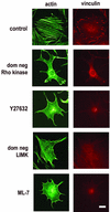
FIG. 1.
FIG. 1.
Inhibition of Rho kinase and its effectors blocks stress fiber formation. hα5-3T3 cells were transiently transfected with a dominant-negative (dom neg) Rho kinase expression vector or dominant-negative LIMK expression vector and then serum starved or else serum starved and then pretreated with 10 μM Y27632 or 10 μM ML-7. Control cells were either transiently transfected with empty vector or pretreated with DMSO. The cells were plated at subconfluence on fibronectin-coated dishes containing coverslips and stimulated with 10 ng of bFGF/ml. Coverslips were collected at 9 h, fixed, permeabilized, and analyzed for f-actin and vinculin by fluorescence microscopy. Bar, 5 μm.
FIG. 2.
FIG. 2.
Enforced clustering of α5β1 integrin rescues sustained ERK activity and mid-G1 phase cyclin D1 expression. (A) hα5-3T3 cells were either transiently transfected (with empty vector, dominant-negative Rho kinase, or dominant-negative LIMK and then serum starved) or serum starved prior to treatment with Y27632. The starved cells (lanes 0) were treated with trypsin, plated on dishes coated with fibronectin or anti-α5β1, and stimulated with 10 ng of bFGF/ml. (B) Starved hα5-3T3 cells were treated with trypsin, held in suspension for 1 h (lanes S), and pretreated with DMSO or ML-7 before being plated on dishes coated with fibronectin or anti-α5β1. Collected cells were lysed and analyzed by immunoblotting with antibodies to pY397 FAK, FAK, cyclin D1, and cdk4 (loading control). ERK activation was determined by gel-shift and by direct detection of dually phosphorylated ERKs (pERK).
FIG. 3.
FIG. 3.
Cell adhesion and stress fibers are required for sustained MEK activity in growth factor-treated cells. (A) Quiescent hα5-3T3 cells were plated at subconflence on fibronectin (FN)-coated dishes and stimulated with 10 ng of bFGF/ml. These cells were treated with DMSO or U0126 30 min before collection, at each of the indicated times. (B) hα5-3T3 cells were transiently transfected with constitutively activated MEK, serum starved, pretreated with DMSO or ML-7, plated on fibronectin or BSA-coated dishes (suspension culture) and stimulated with 10 ng of bFGF/ml. Cells were treated with U0126 or DMSO 5 min prior to the indicated collection time (3 or 9 h). In panels A and B, cell lysates were analyzed for activation of ERK by gel-shift and by direct detection of dually phosphorylated ERKs (pERK). (C) Quiescent hα5-3T3 cells were plated on dishes coated with fibronectin or poly-l-lysine (PLL) and treated with 10 ng of bFGF/ml. At the indicated times, cells were collected and lysed. ERK activation was determined by immunoblotting as described above. Cell lysates were also incubated with anti-ERK or anti-MEK, and the collected immunoprecipitates were used to assess in vitro ERK or MEK activities by phosphorylation of myelin basic protein (MBP) or kinase-dead ERK [GST-ERK (K52R)], respectively. The amounts of ERK or MEK in the immunoprecipitates (IP) were determined by immunoblotting (IB) with anti-ERK or anti-MEK, respectively. Both kinase-dead ERK and MBP remained unphosphorylated when the lysates were incubated with a control serum (not shown). (D) Quiescent hα5-3T3 cells were pretreated with DMSO or ML-7, plated on dishes coated with either fibronectin (FN) or anti-α5β1, and stimulated with 10 ng of bFGF/ml. Cells were collected at the indicated times, lysed, and analyzed for the activation of ERK and MEK activity as described for panel C.
FIG. 4.
FIG. 4.
Integrin signaling and stress fibers act upstream of Ras to sustain ERK activity. (A) Quiescent hα5-3T3 cells were pretreated with DMSO or ML-7, plated on dishes coated with either fibronectin or poly-l-lysine (PLL), and stimulated with 10 ng of bFGF/ml. Cells were collected at the indicated times, lysed, and analyzed for total and GTP-loaded Ras. Identically treated cells were analyzed for Raf-1 activity and the level of total Raf-1 (see Materials and Methods). Total cell lysates were also used to monitor the activities of MEK and ERK by immunoblotting for phosphorylation of MEK at S222 and dual phosphorylation of pERK, respectively. (B) hα5-3T3 cells were transiently transfected with empty vector or FRNK, serum starved for 24 h (0), and then held in suspension for 1 h (lanes S), prior to stimulation on fibronectin (FN)-coated dishes with 10 ng of bFGF/ml. Collected cells were lysed and analyzed by immunoblotting with antibodies to pY397 FAK, FAK, pERK, ERK, cyclin D1, and cdk4 (loading control).
FIG. 5.
FIG. 5.
Distinct effects of Rho kinase inhibition and MLCK inhibition on progression through G1 phase. MEFs were serum starved for 36 h and then pretreated with 10 μM Y27632, 10 μM ML-7, or DMSO. Alternatively, MEFs were transiently transfected with a dominant-negative (dom neg) Rho kinase expression vector or empty vector and then serum starved for 36 h. The cells were plated in dishes containing coverslips and stimulated with 10% FBS in the presence of BrdU. Coverslips were collected and fixed at the indicated times for an analysis of S phase entry by BrdU incorporation, counting ∼150 cells per sample.
FIG. 6.
FIG. 6.
Rac/Cdc42-dependent cyclin D1 expression leads to activation of the G1 phase cdks in the absence of stress fibers. (A) MEFs were serum starved for 48 h; pretreated with DMSO, 10 μM ML-7, or 10 μM Y27632; plated at subconfluence; and stimulated with 10% FBS. At the times shown, collected cells were lysed and equal amounts of total protein were analyzed by immunoblotting for the levels and activation of ERK by using anti-ERK and anti-pERK, respectively. Duplicate filters were analyzed by immunoblotting for the expression of cyclin D1, cdk4, cyclin E, cdk2, p27kip1, and p21cip1. (B) MEFs transiently transfected with empty vector or the dominant-negative (dom neg) Rho kinase expression vector, serum starved for 48 h, plated at subconfluence, and stimulated with 10% FBS. Collected cells were lysed and analyzed by immunoblotting with antibodies against ERK, pERK, cyclin D1, cdk4, and myc (the epitope tag for dom neg Rho kinase). Cell lysates were also incubated with anti-cdk4, and the collected immunoprecipitates were used to assess in vitro cdk4 activity by phosphorylation of GST-Rb. The amount of immunoprecipitated (IP) cdk4 was assessed by immunoblotting (IB) by using filters from the kinase assay. (C) MEFs were transiently transfected with empty vector, serum starved for 48 h, and preincubated with DMSO or U0126 in the absence and presence of Y27632. Alternatively, MEFs were transiently transfected with the PBD expression vector, serum starved for 48 h, and preincubated with DMSO or Y27632. Cell lysates were analyzed by immunoblotting with antibodies to ERK, pERK, cyclin D1, cdk4, and GST (epitope tag for the PBD). (D) serum-starved MEFs were pretreated with DMSO, 10 μM Y27632, or 10 μM ML-7. (E) MEFs were transfected with empty vector or dominant-negative Rho kinase prior to 36 h serum starvation. For both panels D and E, the cells were plated at subconfluence and stimulated with 10% FBS. Cell lysates were incubated with anti-cyclin E, and the collected immunoprecipitates (IP) were used to assess in vitro cyclin E-cdk2 kinase activity by phosphorylation of histone H1. The levels of cyclin E-associated cdk2 was determined by immunoblotting (IB) the kinase assay filters with anti-cdk2.
FIG. 7.
FIG. 7.
Ectopic expression of cyclin D1 rescues S phase entry in ML-7-treated cells. MEFs stably expressing tetracycline (tet)-repressible cyclin D1 were grown to confluence in the presence of tetracycline, washed, and serum starved for 48 h in the presence or absence of tetracycline. In the continued presence or absence of tetracycline, the starved cells were trypsinized and pretreated with 10 μM ML-7 (lanes M) or DMSO (lanes D) as described for MEFs in Materials and Methods. The pretreated cells were plated in 100-mm dishes containing coverslips and stimulated with 10% FBS in the presence of BrdU. Coverslips were collected and fixed at 6 h (□) and 21 h (▪) for an analysis of S phase entry by BrdU incorporation, counting ∼150 cells per sample. The results show mean ± the standard deviation. The remaining cells were collected at 21 h, lysed, and analyzed by immunoblotting with antibodies to cyclin D1 and cdk4 (inset).
FIG. 8.
FIG. 8.
Regulation of cyclin D1 expression by Rho kinase in fibroblasts. Rho kinase-dependent stress fiber formation allows for α5β1 integrin clustering. Clustered α5β1 integrin cooperates with RTK signaling upstream of Ras to sustain Ras, Raf, MEK, and ERK activities. These effects lead to mid-G1 phase cyclin D1 expression and can explain why G1 phase cell cycle progression is stress fiber dependent. Rho kinase is also involved in suppressing the early G1 phase expression of cyclin D1 by Rac/Cdc42. Rac/Cdc42 signaling induces cyclin D1 in early G1 phase, and this effect is independent of stress fiber formation. Since stress fibers do not affect the downregulation p21cip1 or p27kip1 (not depicted), G1 phase progression is independent of stress fiber formation if Rac/Cdc42 induces cyclin D1.

Comment in

  • Findings of misconduct in science.
    [No authors listed] [No authors listed] NIH Guide Grants Contracts (Bethesda). 2007 Jul 20:NOT-OD-07-075. NIH Guide Grants Contracts (Bethesda). 2007. PMID: 17647301 Free PMC article. No abstract available.

Similar articles

Cited by

References

    1. Adnane, J., F. A. Bizouarn, Y. Qian, A. D. Hamilton, and S. M. Sebti. 1998. p21WAF1/CIP1 is upregulated by the geranylgeranyltransferase I inhibitor GGTI-298 through a transforming growth factor beta- and Sp1-responsive element: involvement of the small GTPase RhoA. Mol. Cell. Biol. 18:6962-6970. - PMC - PubMed
    1. Agnew, B. J., L. S. Minamide, and J. R. Bamburg. 1995. Reactivation of phosphorylated actin depolymerizing factor and identification of the regulatory site. J. Biol. Chem. 270:17582-17587. - PubMed
    1. Amano, M., K. Chihara, K. Kimura, Y. Fukata, N. Nakamura, Y. Matsuura, and K. Kaibuchi. 1997. Formation of actin stress fibers and focal adhesions enhanced by Rho kinase. Science 275:1308-1311. - PubMed
    1. Amano, M., M. Ito, K. Kimura, Y. Fukata, K. Chihara, T. Nakano, Y. Matsuura, and K. Kaibuchi. 1996. Phosphorylation and activation of myosin by Rho-associated kinase (Rho kinase). J. Biol. Chem. 271:20246-20249. - PubMed
    1. Aplin, A. E., S. M. Short, and R. L. Juliano. 1999. Anchorage-dependent regulation of the mitogen-activated protein kinase cascade by growth factors is supported by a variety of integrin α chains. J. Biol. Chem. 274:31223-31228. - PubMed

Publication types

MeSH terms

Substances