. 2003 Jun;23(12):4344-55.
doi: 10.1128/MCB.23.12.4344-4355.2003.

The putative GTPases Nog1p and Lsg1p are required for 60S ribosomal subunit biogenesis and are localized to the nucleus and cytoplasm, respectively

Affiliations

The putative GTPases Nog1p and Lsg1p are required for 60S ribosomal subunit biogenesis and are localized to the nucleus and cytoplasm, respectively

George Kallstrom et al. Mol Cell Biol. 2003 Jun.

Abstract

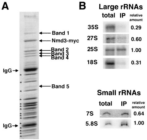
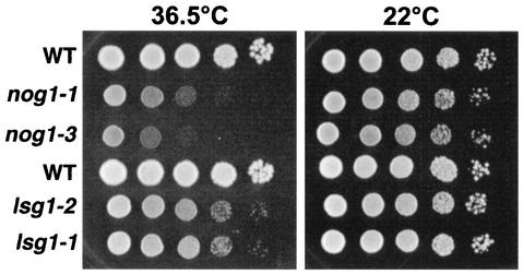
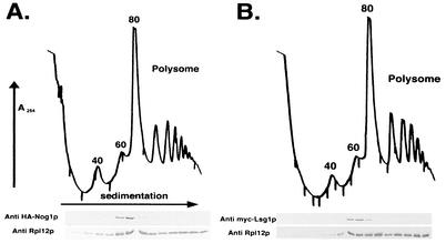
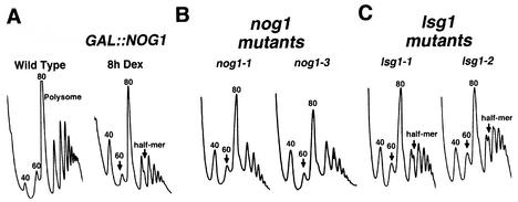
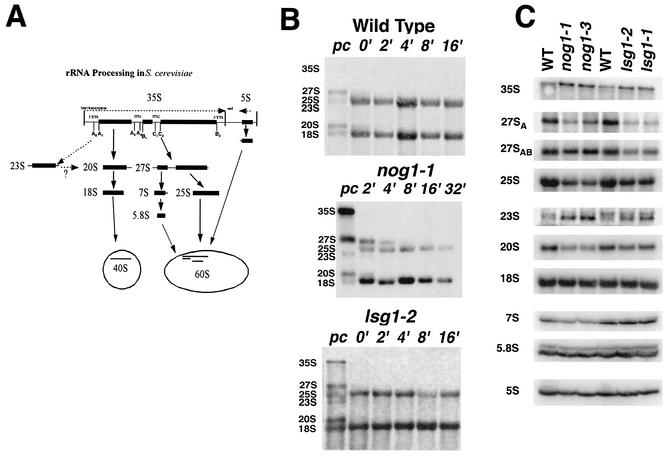
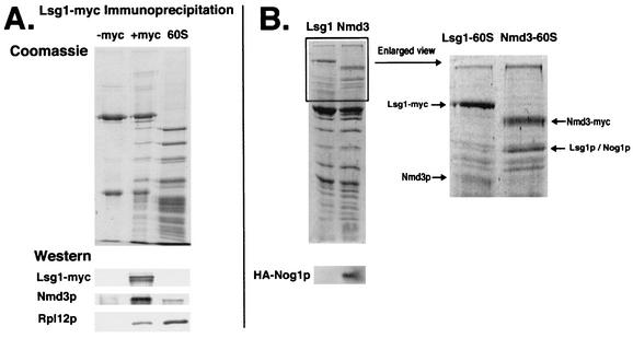
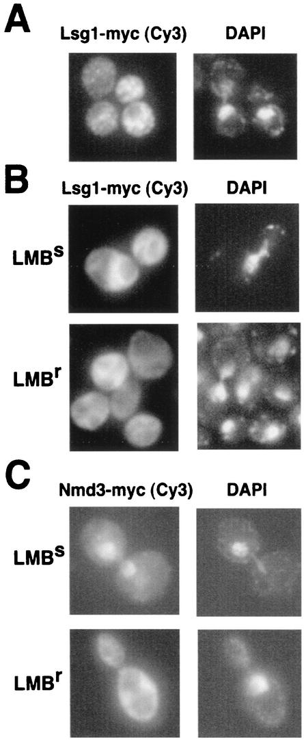
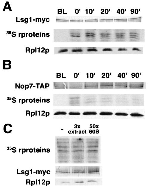
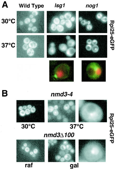
We characterized two essential putative GTPases, Nog1p and Lsg1p, that are found associated with free 60S ribosomal subunits affinity purified with the nuclear export adapter Nmd3p. Nog1p and Lsg1p are nucleolar and cytoplasmic, respectively, and are not simultaneously on the same particle, reflecting the path of Nmd3p shuttling in and out of the nucleus. Conditional mutants of both NOG1 and LSG1 are defective in 60S subunit biogenesis and display diminished levels of 60S subunits at restrictive temperature. Mutants of both genes also accumulate the 60S ribosomal reporter Rpl25-eGFP in the nucleolus, suggesting that both proteins are needed for subunit export from the nucleolus. Since Lsg1p is cytoplasmic, its role in nuclear export is likely to be indirect. We suggest that Lsg1p is needed to recycle an export factor(s) that shuttles from the nucleus associated with the nascent 60S subunit.


Figures

FIG. 1.
FIG. 1.
Immunoprecipitation of Nmd3p-60S complexes. (A) Extracts were prepared from AJY272 (Nmd3-myc) and immunoprecipitated as described previously (19). The proteins in the affinity purified complex were separated by SDS-PAGE (Fig. 1). 60S subunit proteins are indicated by asterisks. Protein not present in the purified 60S sample (labeled in Fig. 1) were excised, digested with trypsin, and analyzed by MALDI-TOF mass spectrometry. (B) RNA was extracted from the immunoprecipitated material prepared as in panel A and compared with total cellular RNA by Northern blotting as described in Materials and Methods. Relative amount indicates the ratio of signal for a given species in the immunoprecipitation (IP) compared to that seen in the total extract when normalized to 25S signals for large RNAs and 5.8S signals for small RNAs.
FIG. 2.
FIG. 2.
Growth of temperature-sensitive mutants on plates. Tenfold serial dilutions of saturated cultures were spotted onto YPD plates and incubated for 3 days at the indicated temperatures. The strains tested were as follows: AJY1124 (GAL1::NOG1) containing plasmid pAJ290 (WT, first row), pAJ633 (nog1-1), or pAJ637 (nog1-3) and AJY1167 (LSG1::KanMX) containing pAJ289 (WT, fourth row), pAJ740 (lsg1-1), or pAJ741 (lsg1-2).
FIG. 3.
FIG. 3.
Nog1p and Lsg1p cosediment with free 60S subunits. Lysates were prepared in the presence of cycloheximide from strain AJY1124 (GAL1::NOG1) containing pAJ290 (HA-NOG1) (A) and strain AJY1126 (GAL1::LSG1) containing pAJ289 (c-myc-LSG1) (B) and then fractionated on 7 to 47% sucrose gradients by ultracentrifugation. The 40S, 60S, 80S, and polysome peaks are labeled in panel A. Fractions were collected while the absorbance at 254 nm was continuously monitored. Proteins were precipitated with trichloroacetic acid, separated on SDS-polyacrylamide gels, transferred to nitrocellulose membranes, and immunoblotted for ribosomal protein Rpl12p and HA or c-myc as indicated.
FIG. 4.
FIG. 4.
Polysome analysis. Extracts were prepared from mid-log-phase cultures and sedimented through 7 to 47% sucrose gradients. Gradients were analyzed by monitoring the absorbance at 254 nm. Arrows indicate reduced free 60S peaks. (A) Wild-type (CH1305, WT) and the glucose-repressible (AJY1118, GAL::NOG1) strains were cultured in galactose-containing medium. Glucose was added to 2%, and the cells were harvested after an additional 8 h. (B) AJY1124 (GAL1::NOG1) containing pAJ633 (nog1-1) or pAJ637 (nog1-3) were grown to log phase at 30°C, followed by incubation at a restrictive temperature (37°C) for 4 h. (C) AJY1167 (LSG1::KanMX) containing pAJ740 (lsg1-1) or pAJ741 (lsg1-2) were cultured as described for the nog1 mutants in panel B.
FIG. 5.
FIG. 5.
rRNA Analysis. (A) Simplified schematic view of rRNA processing in S. cerevisiae. (B) [methyl-3H]methionine pulse-labeling of rRNA was carried out as previously described (18). Wild-type (CH1305), nog1-1 (AJY1124 containing pAJ633), and lsg1-2 (AJY1167 containing pAJ741 (lsg1-2) strains were grown to mid-log phase in selective medium lacking methionine at 30°C. Cells were shifted to 37°C for 2 h and labeled with l-[methyl-3H]methionine for 4 min and then chased with excess unlabeled methionine. Aliquots were collected and frozen 2 min before the addition of chase (lanes pc), immediately after the addition of chase (lanes 0′), and at the indicated times after the addition of chase. Total RNA was prepared and separated on formaldehyde-agarose gels. RNAs were transferred to nylon membranes, sprayed with En3Hance (Dupont) and exposed to X-ray film. (C) Northern blotting of pre- and mature rRNAs. The strains tested were as follows: left three lanes, AJY1124 containing pAJ290 (WT), pAJ633 (nog1-1), or pAJ637 (nog1-3); right three lanes, AJY1167 containing pAJ289 (WT), pAJ741 (lsg1-2), or pAJ740 (lsg1-1). Cultures were grown to early log phase at 26°C, followed by 3 h of incubation at 37°C. RNA was prepared and analyzed by Northern blotting as described in Materials and Methods.
FIG. 6.
FIG. 6.
Coimmunoprecipitation of free 60S by Lsg1p. (A) Extracts from strain CH1305 (wild-type) with pRS416 (−myc) and pAJ901 (+myc) were incubated with monoclonal antibody 9e10 (anti-myc) antibody (Covance) and protein A-beads. Precipitated proteins were eluted from the beads by heating in 1× Laemmli sample buffer and separated by SDS-PAGE. Purified 60S subunit proteins were used for comparison. Proteins were stained with Coomassie blue or transferred to nitrocellulose. Western blotting was performed against Lsg1-myc, Nmd3p, and Rpl12p to assay their presence on the Lsg1p-bound 60S subunit. (B) Extracts from strain AJY272 (NMD3-myc) containing pAJ625 (HA-NOG1) and strain CH1305 containing pAJ901 (LSG1-myc) and pAJ625 were subjected to immunoprecipitation as described in panel A. Precipitated proteins were separated by SDS-PAGE and stained with Coomassie blue or transferred to nitrocellulose for Western blotting for HA-Nog1p.
FIG. 7.
FIG. 7.
Lsg1p is cytoplasmic and does not shuttle in a Crm1-dependent fashion. (A) Strain CH1305 (wild-type) containing pAJ901 (LSG1-myc) was grown to mid-log phase, and immunofluorescence analysis was performed as described in Materials and Methods. (B) Strains MNY7 (LMB resistant [LMBr]) and MNY8 (LMB sensitive [LMBs]) were transformed with pAJ901. (C) As a control, the LMB-dependent nuclear accumulation of Nmd3-myc was monitored in MNY7 and MNY8 containing pAJ538 (NMD3-myc) (20). Strains were cultured and treated with LMB as described in Materials and Methods.
FIG. 8.
FIG. 8.
Time course of labeled subunit binding to Lsg1p in vivo. Cultures of CH1305 containing pAJ901 (LSG1-myc) (A) and NOP7-TAP (B) were pulse-labeled with l-[35S]methionine for 5 min as described in Materials and Methods. At the indicated time points aliquots were removed, extracts prepared, and tagged proteins were affinity purified by using anti-myc antibodies for Lsg1-myc or as described previously for NOP7-TAP (17). Precipitated complexes were subjected to SDS-PAGE and dried for autoradiography or transferred to nitrocellulose for Western blotting. (C) Extracts were prepared from 35S-labeled CH1305 containing pAJ901. Extracts were incubated with a 3-fold excess of unlabeled wild-type extract or with a 50-fold excess of purified unlabeled 60S. Lsg1-myc and associated 60S proteins were immunoprecipitated and analyzed as described in panel A.
FIG. 9.
FIG. 9.
Rpl25-eGFP localization. (A) Rpl25-eGFP was expressed in strain CH1305 (wild-type), strain AJY1124 (GAL1::NOG1) carrying pAJ637 (nog1-3), and strain AJY1167 (LSG1::KanMX) carrying pAJ740 (lsg1-1). Fresh overnight cultures were diluted twofold into fresh medium. After 30 min at 30°C, each culture was divided, and one half of each shifted to 37°C for 3 h. Enlarged examples compare the localization of the Rpl25-eGFP with that of DNA stained with Hoechst 34442. For the colocalization experiments, the strains were AJY1124 (GAL1::NOG1) carrying pAJ633 (nog1-1) and AJY1167 (LSG1::KanMX) carrying pAJ740 (lsg1-1). GFP fluorescence (green) and Hoechst fluorescence (red) were visualized in separate channels, artificially colored, and merged. (B) Rpl25-eGFP was expressed in AJY734 (nmd3-4) at a permissive (30°C) or a nonpermissive (37°C) temperature for 3 h or coexpressed in CH1305 containing pAJ368 (GAL1::NMD3Δ100) (20) in the presence of the inducer galactose (gal) or in the presence of the noninducing sugar raffinose (raf). Enlarged examples of cells are shown.
FIG. 10.
FIG. 10.
Simplified diagram of Nmd3p and Lsg1p binding to 60S subunits. Nmd3p loads onto the pre-60S subunit in the nucleus, possibly as the nascent subunit emerges from the nucleolus. Nmd3p remains associated with the subunit during export to the cytoplasm. Tif6p, Arx1p, Crm1(Xpo1p), and Ran(Gsp1p) also accompany the subunit during export but are not shown for clarity. In the cytoplasm, Lsg1p likely binds to the newly exported free 60S subunit, and we suggest that it is required for recycling an as-yet-unidentified factor to the nucleus. Both Nmd3p and Lsg1p are released prior to or upon subunit joining. When released, Nmd3p can reenter to the nucleus for another round of subunit export or bind to a recycling free 60S subunit after translation termination. Lsg1p, on the other hand, is restricted to the cytoplasm and, after it is released from a subunit, it can bind to a newly exported 60S subunit or to a recycling free 60S subunit as with Nmd3p.

Similar articles

Cited by

References

    1. Bassler, J., P. Grandi, O. Gadal, T. Lessmann, E. Petfalski, D. Tollervey, J. Lechner, and E. Hurt. 2001. Identification of a 60S preribosomal particle that is closely linked to nuclear export. Mol. Cell 8:517-529. - PubMed
    1. Basu, U., K. Si, J. R. Warner, and U. Maitra. 2001. The Saccharomyces cerevisiae TIF6 gene encoding translation initiation factor 6 is required for 60S ribosomal subunit biogenesis. Mol. Cell. Biol. 21:1453-1462. - PMC - PubMed
    1. Bécam, A. M., F. Nasr, W. J. Racki, M. Zagulski, and C. J. Herbert. 2001. Ria1p (Ynl163c), a protein similar to elongation factors 2, is involved in the biogenesis of the 60S subunit of the ribosome in Saccharomyces cerevisiae. Mol. Gen. Genet. 266:454-462. - PubMed
    1. Belk, J. P., F. He, and A. Jacobson. 1999. Overexpression of truncated Nmd3p inhibits protein synthesis in yeast. RNA 5:1055-1070. - PMC - PubMed
    1. Dragon, F., J. E. G. Gallagher, P. A. Compagnone-Post, B. M. Mitchell, K. A. Porwancher, K. A. Wehner, S. Wormsley, R. E. Settlage, J. Shabanowitz, Y. Osheim, A. L. Beyer, D. F. Hunt, and S. J. Baserga. 2002. A large nucleolar U3 ribonucleoprotein required for 18S ribosomal RNA biogenesis. Nature 417:967-970. - PMC - PubMed

Publication types

MeSH terms