

Competition and representation during memory retrieval: roles of the prefrontal cortex and the posterior parietal cortex
- PMID: 12773617
- PMCID: PMC165889
- DOI: 10.1073/pnas.0832374100
Competition and representation during memory retrieval: roles of the prefrontal cortex and the posterior parietal cortex
Abstract
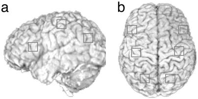
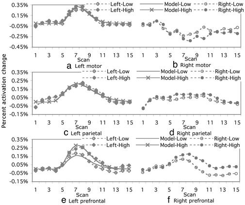
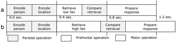
In this functional-MRI study we examined the hypothesis that the prefrontal cortex responds differently to the extent of competition during retrieval, whereas the parietal cortex is responsible for problem representation that should not be directly related to the competition. Participants mastered arbitrary person-location pairs, and their recognition memory was tested in a functional-MRI session. The pairs were constructed such that a person was associated with one, two, or three different locations and vice versa. The recognition time increased with the number of associations, reflecting increased competition. A confirmatory analysis of imaging data with prespecified prefrontal and parietal regions showed that, although both regions were highly involved during memory retrieval, only the prefrontal region responded to the levels of competition. This result was consistent with predictions of an information-processing model as well as with an exploratory identification of regions of interest.
Figures
Similar articles
-
Role of prefrontal and parietal cortices in associative learning.Cereb Cortex. 2008 Apr;18(4):904-14. doi: 10.1093/cercor/bhm123. Epub 2007 Aug 3. Cereb Cortex. 2008. PMID: 17675369 Free PMC article.
-
An information-processing model of three cortical regions: evidence in episodic memory retrieval.Neuroimage. 2005 Mar;25(1):21-33. doi: 10.1016/j.neuroimage.2004.11.001. Epub 2005 Jan 25. Neuroimage. 2005. PMID: 15734340
-
Functional asymmetry of human prefrontal cortex in verbal and non-verbal episodic memory as revealed by fMRI.Brain Res Cogn Brain Res. 2000 Jan;9(1):73-83. doi: 10.1016/s0926-6410(99)00047-6. Brain Res Cogn Brain Res. 2000. PMID: 10666559
-
The neural basis of episodic memory: evidence from functional neuroimaging.Philos Trans R Soc Lond B Biol Sci. 2002 Aug 29;357(1424):1097-110. doi: 10.1098/rstb.2002.1102. Philos Trans R Soc Lond B Biol Sci. 2002. PMID: 12217177 Free PMC article. Review.
-
Top-down and bottom-up attention to memory: a hypothesis (AtoM) on the role of the posterior parietal cortex in memory retrieval.Neuropsychologia. 2008;46(7):1828-51. doi: 10.1016/j.neuropsychologia.2008.03.022. Epub 2008 Apr 8. Neuropsychologia. 2008. PMID: 18471837 Review.
Cited by
-
Anticipation of conflict monitoring in the anterior cingulate cortex and the prefrontal cortex.Proc Natl Acad Sci U S A. 2007 Jun 19;104(25):10330-4. doi: 10.1073/pnas.0703225104. Epub 2007 Jun 11. Proc Natl Acad Sci U S A. 2007. PMID: 17563353 Free PMC article.
-
Overcoming suppression in order to remember: contributions from anterior cingulate and ventrolateral prefrontal cortex.Cogn Affect Behav Neurosci. 2008 Jun;8(2):211-21. doi: 10.3758/cabn.8.2.211. Cogn Affect Behav Neurosci. 2008. PMID: 18589510 Free PMC article.
-
Role of prefrontal and parietal cortices in associative learning.Cereb Cortex. 2008 Apr;18(4):904-14. doi: 10.1093/cercor/bhm123. Epub 2007 Aug 3. Cereb Cortex. 2008. PMID: 17675369 Free PMC article.
-
Distinct roles of the anterior cingulate and prefrontal cortex in the acquisition and performance of a cognitive skill.Proc Natl Acad Sci U S A. 2006 Aug 22;103(34):12941-6. doi: 10.1073/pnas.0605493103. Epub 2006 Aug 16. Proc Natl Acad Sci U S A. 2006. PMID: 16914528 Free PMC article.
-
Conditional routing of information to the cortex: a model of the basal ganglia's role in cognitive coordination.Psychol Rev. 2010 Apr;117(2):541-74. doi: 10.1037/a0019077. Psychol Rev. 2010. PMID: 20438237 Free PMC article.
References
-
- Collins, A. M. & Quillian, M. R. (1969) J. Verbal Learn. Verbal Behav. 8, 240-247.
-
- Cabeza, R., Dolcos, F., Graham, R. & Nyberg, L. (2002) Neuroimage 16, 317-330. - PubMed
-
- Dove, A., Pollmann, S., Schubert, T., Wiggins, C. J. & von Cramon, D. Y. (2000) Cognit. Brain Res. 9, 103-109. - PubMed
-
- Kimberg, D. Y., Aquirre, G. K. & D'Esposito, M. (2000) Cognit. Brain Res. 10, 189-196. - PubMed
Publication types
MeSH terms
Substances
Grants and funding
LinkOut - more resources
Full Text Sources
Medical
