. 2003 Jun 10;100(12):7366-71.
doi: 10.1073/pnas.1232342100. Epub 2003 May 28.

The hSK4 (KCNN4) isoform is the Ca2+-activated K+ channel (Gardos channel) in human red blood cells

Affiliations

The hSK4 (KCNN4) isoform is the Ca2+-activated K+ channel (Gardos channel) in human red blood cells

Joseph F Hoffman et al. Proc Natl Acad Sci U S A. .

Abstract

The question is, does the isoform hSK4, also designated KCNN4, represent the small conductance, Ca2+-activated K+ channel (Gardos channel) in human red blood cells? We have analyzed human reticulocyte RNA by RT-PCR, and, of the four isoforms of SK channels known, only SK4 was found. Northern blot analysis of purified and synchronously growing human erythroid progenitor cells, differentiating from erythroblasts to reticulocytes, again showed only the presence of SK4. Western blot analysis, with an anti-SK4 antibody, showed that human erythroid progenitor cells and, importantly, mature human red blood cell ghost membranes, both expressed the SK4 protein. The Gardos channel is known to turn on, given inside Ca2+, in the presence but not the absence of external Ko+ and remains refractory to Ko+ added after exposure to inside Ca2+. Heterologously expressed SK4, but not SK3, also shows this behavior. In inside-out patches of red cell membranes, the open probability (Po) of the Gardos channel is markedly reduced when the temperature is raised from 27 to 37 degrees C. Net K+ efflux of intact red cells is also reduced by increasing temperature, as are the Po values of inside-out patches of Chinese hamster ovary cells expressing SK4 (but not SK3). Thus the envelope of evidence indicates that SK4 is the gene that codes for the Gardos channel in human red blood cells. This channel is important pathophysiologically, because it represents the major pathway for cell shrinkage via KCl and water loss that occurs in sickle cell disease.


Figures

Fig. 1.
Fig. 1.
The SK4 isoform of the Ca2+-activated K+ channel is present in human reticulocytes. PCR products were obtained with the isoform-specific primers defined in Table 1. Single-stranded cDNA derived from reticulocytes (see Methods) was used as template. Lanes 1 and 2 had, respectively, 25- and 4-μl samples applied to the gel. Lane 3 is a water control. The mass ladder (in base pairs) is shown at left (Life Technologies, Grand Island, NY). The expected product size was 1,767, and a product of approximately this size was found in lanes 1 and 2. The product was sequenced in this and other analyses and was shown to have >98% identify with the expected sequence for SK4.
Fig. 2.
Fig. 2.
Northern blots probed for the mRNA encoding the Ca2+-activated K+ channel isoform, SK4, using RNA prepared from cultured human erythroid progenitor cells at different stages of maturation (see Methods for details). Also shown is the positive control blot for SK4 (Right), where H is heart; B, brain; P, placenta; L, lung; Li, liver; S, skeletal muscle; K, kidney; and Pa, pancreas, all from human mRNA. These results parallel our previous finding that SK4 is present in reticulocytes. Note that SK4 is present in the progenitor cells at day 7 increasing with differentiation through day 12. The transcript size is indicated on the ordinate.
Fig. 3.
Fig. 3.
Western blots showing that the protein for the Gardos channel isoform, SK4, is present in cultured human erythroid progenitor cells and in ghost membranes made from mature human red blood cells. The antibody was prepared against an SK4-specific peptide and used as described in Methods. The two positive controls are human parotid gland (P) and kidney (K) with the negative control being brain (B). It is clear that a band of the appropriate molecular weight is present in the human erythroid progenitor cells as they mature from days 7 to 13. It is also evident that the SK4 band is present in human red cell ghost membranes (RBC). The decrease in the blot intensity of the D13 band compared with D7 is primarily due to the decreased protein content (cell number) of cells loaded onto the gel. The slight variation in the molecular weights of SK4 bands seen in the progenitor cells, relative to the other bands, may be due to posttranslational modification or higher salt concentration in the loading mixture. It should also be mentioned that, except in the parotid lane, there are higher molecular weight bands (not shown) that in each case react with the antibody. Importantly, preincubation of the antibody with purified peptide that contains the antigenic epitope produces a complete loss of reactivity in all lanes except in brain, where it is much reduced, and in ghosts, where it is only faintly present in the highest molecular weight bands (data not shown).
Fig. 4.
Fig. 4.
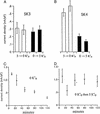
The effect of external formula image on the activity of heterologously expressed SK3 and SK4 channels in CHO cells. Cells were washed twice in 0 mM formula image and then incubated with 100 μM 1,2-bis(2-aminophenoxy)ethane-N,N,N,N′-tetraacetate or -tetraacetic acid–acetoxymethyl ester (BAPTA-AM) in either 5 mM formula image (open bars) or 0 mM formula image (dark bars) containing media for 3–5 h at room temperature (see Methods). At the end of this period, cells were perfused with incubation solution before and during seal formation, breakthrough, and achievement of whole-cell recording mode. Current amplitude at 60 mV was then measured for each cell first during continued perfusion with incubation solution and then after switching from 0 to 5 formula image or vice versa. The whole-cell currents in expressed SK3 channels (A) were insensitive to the presence or absence of formula image, whereas the currents in expressed SK4 channels (B) were markedly diminished by preincubation in 0 mM formula image. (C) The decrease in whole-cell currents that occurred during the first 2 h of incubation of SK4 expressing cells in 0 mM formula image.(D) SK4 cells can recover from incubation in 0 mM formula image by reexposure to 5 mM formula image over this time period. SK4 cells exposed for longer periods of time (B, dark bars) to 0 mM formula image remain refractory to 5 mM formula image. Error bars represent ± SEM, where n = 9–15 separate observations with SK3 (A) and 11–15 for SK4 (B); in C and D, n = 3–12.
Fig. 5.
Fig. 5.
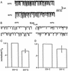
The sensitivity of hSK4 channels to changes in temperature. Results of single-channel recordings from inside-out patches excised from CHO cells stably expressing hSK4 are presented. The bathing solution contained 1 μM free Ca2+ with 130 mM K+ on both sides of the patch (see Methods). Recordings were performed at -80 mV first at 25°C and then, within 10 s, at 35°C. The single-channel activity is shown in A and on an expanded time scale in B. The bars labeled 35°Cin C and D represent the normalized values at 25°C (taken as 1.0) of the open probability (Po) of each channel from a given patch. The results presented in C represent control characteristics of SK4 channels, whereas in D, thioredoxin peroxidase (see Methods) has been added to the cytoplasmic bathing medium for reasons explained in the text. The error bars are ± SEM where n = 6 in C and n = 4 in D. Lumping the results of C and D together, the mean difference between the values at 25 compared with 37°C is 0.731 ± 0.072 SEM, with P < 0.05.

References

    1. Joiner, W. J., Wang, L.-Y., Tang, M. D. & Kaczmarek, L. K. (1997) Proc. Natl. Acad. Sci. USA 94, 11013-11018. - PMC - PubMed
    1. Ishii, T. M., Silvia, C., Hirschberg, B., Bond, C. T., Adelman, J. P. & Maylie, J. (1997) Proc. Natl. Acad. Sci. USA 94, 11651-11656. - PMC - PubMed
    1. Logsdon, N. J., Kang, J., Togo, J. A., Christian, E. P. & Aiyar, J. (1997) J. Biol. Chem. 272, 32723-32726. - PubMed
    1. Köhler, M., Hirschberg, B., Bond, C. T., Kinzie, J. M., Marrion, N. V., Maylie, J. & Adelman, J. P. (1996) Science 273, 1709-1714. - PubMed
    1. Warth, R., Hamm, K., Bleich, M., Kunzelmann, K., von Hahn, T., Schreiber, R., Ullrich, E., Mengel, M., Trautmann, N., Kindle P., et al. (1999) Pflügers Arch. 438, 437-444. - PubMed

Publication types

MeSH terms