. 2003 Jun 10;100(12):7021-6.
doi: 10.1073/pnas.1237797100. Epub 2003 May 28.

Cu/Zn superoxide dismutase mutants associated with amyotrophic lateral sclerosis show enhanced formation of aggregates in vitro

Affiliations

Cu/Zn superoxide dismutase mutants associated with amyotrophic lateral sclerosis show enhanced formation of aggregates in vitro

P B Stathopulos et al. Proc Natl Acad Sci U S A. .

Abstract

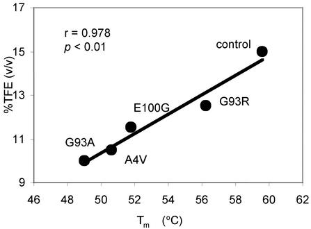
Mutations in Cu/Zn superoxide dismutase (SOD) are associated with the fatal neurodegenerative disorder amyotrophic lateral sclerosis (ALS). There is considerable evidence that mutant SOD has a gain of toxic function; however, the mechanism of this toxicity is not known. We report here that purified SOD forms aggregates in vitro under destabilizing solution conditions by a process involving a transition from small amorphous species to fibrils. The assembly process and the tinctorial and structural properties of the in vitro aggregates resemble those for aggregates observed in vivo. Furthermore, the familial ALS SOD mutations A4V, G93A, G93R, and E100G decrease protein stability, which correlates with an increase in the propensity of the mutants to form aggregates. These mutations also increase the rate of protein unfolding. Our results suggest three possible mechanisms for the increase in aggregation: (i) an increase in the equilibrium population of unfolded or of partially unfolded states, (ii) an increase in the rate of unfolding, and (iii) a decrease in the rate of folding. Our data support the hypothesis that the gain of toxic function for many different familial ALS-associated mutant SODs is a consequence of protein destabilization, which leads to an increase in the formation of cytotoxic protein aggregates.


Figures

Fig. 1.
Fig. 1.
DSC of apo and holo SODs at pH 7.8 (A) and apo SODs at pH 5.4 (B). (A) Specific heat capacity per mol dimer versus temperature for E100G (dashed line) and control SOD (solid line), with reference buffer–buffer scan subtracted. Solutions contained 0.2–0.7 mg/ml protein in 20 mM Hepes, pH 7.8. Apo and holo E100G have lower stability and unfold at lower temperatures than the corresponding forms of the control AS protein. (B) Specific heat capacity per mol dimer versus temperature for (left to right) apo A4V (dashed line), apo E100G (dotted line), and apo control (solid line) with reference buffer–buffer scan and then specific heat capacity of native state subtracted. Solutions contained 0.2 mg/ml protein in 20 mM Mes, pH 5.4. The shapes of the traces are distorted compared with those at pH 7.8 due to exothermic aggregation. Arrowheads show the corresponding onset of aggregation for (left to right) apo A4V, apo E100G, and apo control, as measured by 90° light scattering.
Fig. 2.
Fig. 2.
Rates of unfolding for holo SODs in 6 M GdmCl (• and ▪) and apo SODs in 4 M GdmCl (○ and □) in 20 mM Hepes, pH 7.8, 25°C. Owing to the slower unfolding of AS SOD, both the fast (• and ○) and slow (▪ and □) unfolding phases could be measured for this protein. Because of faster unfolding of the mutants, the faster phase is largely complete in the experimental dead time, and only the slow phase was measured.
Fig. 3.
Fig. 3.
ThT and Congo red binding properties of SOD aggregates. Spectra are shown for solutions with (○) and without (•) protein. (A) ThT fluorescence emission spectra for TFE-induced aggregates of apo G93A. (B) Congo red absorbance spectra for TFE-induced aggregates of apo G93A. (C) ThT fluorescence emission spectrum for heat-induced aggregates of apo G93R. (D) Congo red absorbance spectrum for heat-induced aggregates of apo A4V.
Fig. 4.
Fig. 4.
Correlation between propensity for aggregation and conformational stability of apo SOD. Minimum percentage of TFE (vol/vol) required to induce protein aggregation monitored by enhancement of ThT fluorescence versus Tm. Aggregation was induced by adding protein to a final concentration of 0.2 mg/ml in 50 mM acetate buffer, pH 5.5, at 25°C. The data fit to a straight line by least-squares regression have a high correlation coefficient (r = 0.978) and a low probability of chance correlation (P < 0.01).
Fig. 5.
Fig. 5.
Electron microscopy of in vitro SOD aggregates induced by TFE (A–C) and heat (D–F) showing representative aggregate morphologies. Bars are 100 nm. (A) Amorphous aggregates (right middle), as well as fibrils associated with amorphous aggregates (middle) and merging into larger heavy-staining aggregates (left). (B) Single fibril of diameter ≈12–15 nm (right arrow) apparently consisting of two narrower strands loosely wound together. Single fibrils associate in pairs (left arrow) that are aligned or loosely twisted together to form thicker fibrils with net diameter of ≈15–25 nm. (C) Large, heavily stained amorphous fractal-like aggregates. (D) Amorphous light-colored aggregate spheres or distorted spheres up to ≈100 nm in diameter, associated with thinner fibrils. (E) Higher magnification of D shows heat-induced fibrils resembling TFE-induced fibrils (B). (F) Mechanism of aggregate assembly whereby amorphous light-colored aggregate spheres of various diameters elongate into thinner fibrils. (A–E) Apo G93A. (F) Apo A4V.
Fig. 6.
Fig. 6.
Heat-induced aggregation of apo SOD monitored by 90° light scattering. From left to right, apo G93A (▵), apo A4V (•), apo E100G (□), and apo control (⋄) are shown. For clarity, absolute signals have different offsets added. Solutions contained 20 mM Mes, pH 5.4, and 0.010–0.025 mg/ml protein.

References

    1. Fridovich, I. (1986) Adv. Enzymol. Relat. Areas Mol. Biol. 58, 61–97. - PubMed
    1. Lepock, J. R., Frey, H. E. & Hallewell, R. A. (1990) J. Biol. Chem. 265, 21612–21618. - PubMed
    1. Parge, H. E., Hallewell, R. A. & Tainer, J. A. (1992) Proc. Natl. Acad. Sci. USA 89, 6109–6113. - PMC - PubMed
    1. Rosen, D. R., Siddique, T., Patterson, D., Figlewicz, D. A., Sapp, P., Hentati, A., Donaldson, D., Goto, J., O'Regan, J. P., Deng, H. X., et al. (1993) Nature 362, 59–62. - PubMed
    1. Rowland, L. P. & Shneider, N. A. (2001) N. Engl. J. Med. 344, 1688–1700. - PubMed

Publication types

MeSH terms