Review
. 2003 Jun;4(6):565-70.
doi: 10.1038/sj.embor.embor869.

Getting a grip on non-native proteins

Affiliations
Review

Getting a grip on non-native proteins

Peter C Stirling et al. EMBO Rep. 2003 Jun.

Abstract

It is an underappreciated fact that non-native polypeptides are prevalent in the cellular environment. Native proteins have the folded structure, assembled state and cellular localization required for activity. By contrast, non-native proteins lack function and are particularly prone to aggregation because hydrophobic residues that are normally buried are exposed on their surfaces. These unstable entities include polypeptides that are undergoing synthesis, transport to and translocation across membranes, and those that are unfolded before degradation. Non-native proteins are normal, biologically relevant components of a healthy cell, except in cases in which their misfolding results from disease-causing mutations or adverse extrinsic factors. Here, we explore the nature and occurrence of non-native proteins, and describe the diverse families of molecular chaperones and coordinated cellular responses that have evolved to prevent their misfolding and aggregation, thereby maintaining quality control over these potentially damaging protein species.


Figures
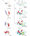
Figure 1
Figure 1
Folding pathways of engrailed homeodomain and egg-white lysozyme. (A) The engrailed homeodomain (En-HD) folds rapidly, in nanoseconds to microseconds (Mayor et al., 2003). (B) Lysozyme has two significantly populated intermediates and folds more slowly, with a timescale of milliseconds in vitro. The majority (70%) of the lysozyme protein population folds relatively quickly into the α-domain intermediate, but is slow to reach the near-native, short-lived αβ-intermediate. Another 20% rapidly forms the αβ-intermediate directly. The α-domain is shown in red, and the β-domain in yellow. Reproduced in part, with permission from Dinner et al., 2000.
Figure 2
Figure 2
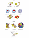
Chaperone structures, grouped according to how they interact with their non-native substrates. (A) The carboxy-terminal substrate-binding domain of Escherichia coli DnaK, comprising a β-sheet cradle and an α-helical lid, bound to a peptide (red; NRLLLTG). (B) Calnexin has a globular lectin domain (yellow) and an extended arm domain (gold). (C,D) Cryoelectron microscopy (cryo-EM) reconstructions of the chaperonin CCT (blue) in complex with (C) actin and (D) tubulin (both in red). (E) HslU AAA ATPase (blue) shown on top of the HslV protease (grey). (F) Archaeal prefoldin (α-subunits shown in yellow; β-subunits shown in gold). (G) Cryo-EM reconstruction of eukaryotic prefoldin (gold) in complex with non-native actin (red). (H) SecB dimer of dimers (the different colours indicate monomers), with the putative substrate-binding groove shown in red. (I) A cofactor-A dimer (yellow and grey) with conserved putative binding residues (red). (J) A small heat-shock protein (Hsp16.5) from Methanococcus jannaschii forms a spherical oligomer; each dimer building-block is shown in a different colour. (K) The periplasmic chaperone PapD (grey) supplies a β-strand (red) to the incomplete PapK immunoglobulin fold (yellow). Structures are not shown to scale. The Protein Database identification numbers for these proteins are as follows: DnaK–peptide, 1DKX; calnexin, 1JHN; HslUV, 1G3I; prefoldin, 1FXK; SecB, 1FX3; cofactor A, 1QSD; sHsp, 1SHS; PapD-K, 1PDK.
Figure 3
Figure 3
An aggresome formed in HeLa cells by overexpression of a green-fluorescent-protein fusion to the polyglutamine region of huntingtin (green), the protein involved in the neurodegenerative disorder, Huntington's disease. The nucleus is shown in blue, and the microtubules in red.
None
None
None

Similar articles

Cited by

References

    1. Chernoff Y.O., Lindquist S.L., Ono B., Inge-Vechtomov S.G. & Liebman S.W. (1995) Role of the chaperone protein Hsp104 in propagation of the yeast prion-like factor [psi+]. Science, 268, 880–884. - PubMed
    1. Clark J.I. & Muchowski P.J. (2000) Small heatshock proteins and their potential role in human disease. Curr. Opin. Struct. Biol., 10, 52–59. - PubMed
    1. Daggett V. & Fersht A.R. (2003) Is there a unifying mechanism for protein folding? Trends Biochem. Sci., 28, 18–25. - PubMed
    1. Dinner A.R., Sali A., Smith L.J., Dobson C.M. & Karplus M. (2000) Understanding protein folding via free-energy surfaces from theory and experiment. Trends Biochem. Sci., 25, 331–339. - PubMed
    1. Farr G.W., Furtak K., Rowland M.B., Ranson N.A., Saibil H.R., Kirchhausen T. & Horwich A.L. (2000) Multivalent binding of nonnative substrate proteins by the chaperonin GroEL. Cell, 100, 561–573. - PubMed

Publication types