. 2003 May;4(5):484-90.
doi: 10.1038/sj.embor.embor821.

Stimulation of c-MYC transcriptional activity and acetylation by recruitment of the cofactor CBP

Affiliations

Stimulation of c-MYC transcriptional activity and acetylation by recruitment of the cofactor CBP

Jörg Vervoorts et al. EMBO Rep. 2003 May.

Abstract

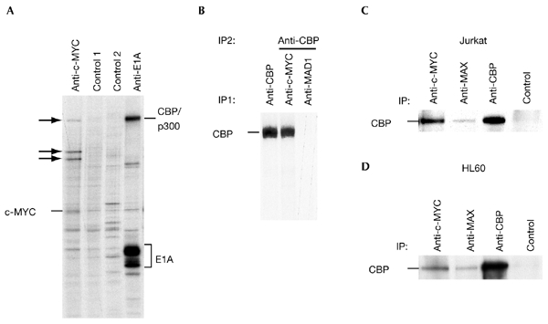
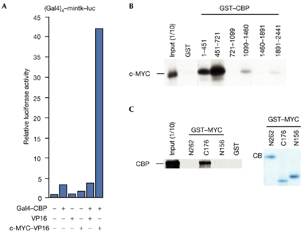
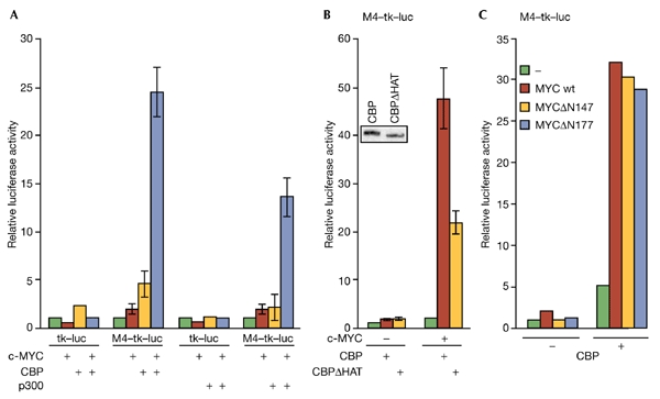
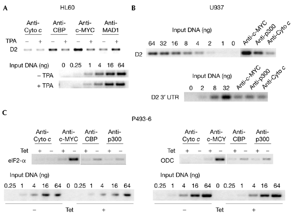
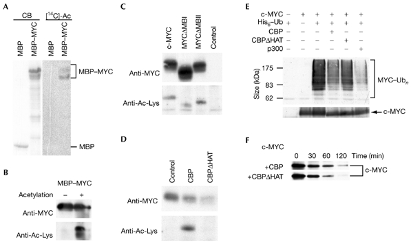
The c-MYC oncoprotein regulates various aspects of cell behaviour by modulating gene expression. Here, we report the identification of the cAMP-response-element-binding protein (CBP) as a novel c-MYC binding partner. The two proteins interact both in vitro and in cells, and CBP binds to the carboxy-terminal region of c-MYC. Importantly, CBP, as well as p300, is associated with E-box-containing promoter regions of genes that are regulated by c-MYC. Furthermore, c-MYC and CBP/p300 function synergistically in the activation of reporter-gene constructs. Thus, CBP and p300 function as positive cofactors for c-MYC. In addition, c-MYC is acetylated in cells. This modification does not require MYC box II, suggesting that it is independent of TRRAP complexes. Instead, CBP acetylates c-MYC in vitro, and co-expression of CBP with c-MYC stimulates in vivo acetylation. Functionally, this results in a decrease in ubiquitination and stabilization of c-MYC proteins. Thus, CBP and p300 are novel functional binding partners of c-MYC.


Figures

Figure 1
Figure 1
Association of endogenous c-MYC and CBP in cells.(A) Human embryonic kidney 293 (HEK293) cells were labelled with [35S]methionine for 2 h before lysis in F buffer. Immunoprecipitations (IPs) were performed with the rat monoclonal antibody 6A10 (specific for c-MYC), protein-G–sepharose beads only (control 1), monoclonal antibody 5C9 (specific for MAD1; control 2) and the mouse monoclonal antibody M73 specific for E1A. The immunoprecipitates were analysed by SDS–polyacrylamide gel electrophoresis (SDS–PAGE). Arrows indicate co-immunoprecipitated proteins. (B) HEK293 cells were treated as in (A). IPs were first performed with C20 anti-cAMP-response-element-binding protein (CBP), monoclonal antibody 6A10 or monoclonal antibody 5C9. Co-immunoprecipitated proteins were eluted in AB buffer and CBP was immunoprecipitated (using the C20 antibody) and analysed by SDS–PAGE. (C) Exponentially growing Jurkat T cells were lysed in F buffer and IPs were performed as indicated using N262 to immunoprecipitate c-MYC, C17 for MAX, AC238 for CBP and SC577 for Gal4, as a control. CBP was then detected by immunoblotting using C20. (D) Co-IPs were performed as in (C), using lysates from promyelocytic HL60 cells.
Figure 2
Figure 2
Interaction of MYC and CBP in overexpression experiments and in vitro. (A) Interaction of c-MYC and CBP (cAMP-response-element-binding protein) detected using a mammalian two-hybrid system. Constructs expressing Gal4–CBP (50 ng), c-MYC–VP16 (2 μg) and VP16 (2 μg) were co-expressed, as indicated, with the Gal4–mintk–luc (a fusion of Gal4, the minimal thymidine-kinase promoter and luciferase) reporter gene construct (2 μg) in U2OS cells. (B) Glutathione-S-transferase (GST)–CBP fusion proteins (1–2 μg) were used to detect interactions with in vitro transcribed and translated c-MYC. One-tenth of the in vitro transcribed/translated material was loaded as a control (input (1/10)). (C) Binding of in vitro transcribed and translated CBP to GST–MYC fusion proteins was analysed as indicated (left panel). A Coomassie-blue (CB)-stained gel containing the three GST–MYC fusion proteins is shown (right panel).
Figure 3
Figure 3
CBP stimulates c-MYC-dependent transactivation and is recruited to MYC-regulated promoters. (A) Reporter gene constructs with (M4–tk–luc; 2 μg) or without (tk–luc; 2 μg) four MYC/MAX binding sites (M4) were co-transfected with expression plasmids for c-MYC (1 μg), Gal4–CBP (5 μg) and Gal4–p300 (5 μg) into U2OS cells as indicated. (B) Transient transfections were performed as in (A). The inset shows the expression of Gal4–CBP and Gal4–CBPΔHAT, as determined by immunoblotting using the SC577 antibody. (C) Transient transfections were performed as in (A). In MYCΔN147 and MYCΔN177, the transactivation domain is deleted. CB,. cAMP-response-element-binding protein; Luc, luciferase; tk, thymidine kinase promoter; wt, wild type.
Figure 4
Figure 4
CBP and p300 are recruited to MYC target genes. (A) HL60 cells were grown exponentially (−) or induced to differentiate with 12-O-tetra-decanoyl-phorbol 13-acetate (TPA) (+) for 12 h. Chromatin immunoprecipitations were performed with antibodies recognizing the indicated proteins (c-MYC, N262; MAD1, C20; CBP, A22; Cyto c, SC7159). For PCR reactions, primers that amplify the region of the cyclin D2 promoter that contains the MYC/MAX binding site were used. For comparison, serial dilutions of input chromatin DNA were analysed. (B) Chromatin immunoprecipitations were performed on lysates of exponentially growing U937 cells. Specific antibodies were used as indicated (c-MYC, N262; p300, C22; Cyto c, SC7159). PCR reactions were carried out as in (A). (C) P493-6 cells were released from a tetracycline (Tet) block (−) or were mock treated (+). Cells were then crosslinked and processed. Immunoprecipitations and PCR reactions were carried out as in (A) and (B). CBP, cAMP-response-element-binding protein; D2, cyclin D2; ODC, ornithine decarboxylase; UTR, untranslated region.
Figure 5
Figure 5
c-MYC is acetylated by CBP. (A) Maltose-binding protein (MBP)–MYC and MBP were incubated with His–CBP in the presence of [14C]acetyl-coenzyme A (acetyl-CoA). A Coomassie-blue-stained gel (CB) and the corresponding autoradiograph ([14C]Ac) are shown. (B) MBP–MYC was acetylated in the presence of His–CBP and acetyl-CoA or was mock-treated. Proteins were detected by western blotting using a MYC-specific antibody (9E10) or acetyl-lysine (Ac-Lys)-specific antibodies (upper and lower panels, respectively). (C) FLAG-tagged c-MYC and MYC mutants were expressed transiently in COS7 cells (10 μg of each construct), immunoprecipitated from F-buffer lysates using FLAG-specific antibodies and analysed by western blotting using monoclonal antibody 9E10 (upper panel) or Ac-Lys-specific antibodies (lower panel). (D) FLAG-tagged c-MYC was co-expressed in COS7 cells with Gal4–CBP, Gal4–CBPΔHAT or vector only. Immunoprecipitations and western blot analyses were performed as in (C). (E) The proteins indicated were expressed in human embryonic kidney (HEK) 293 cells and purified over Talon beads, and MYC proteins were identified using monoclonal antibody 9E10 (upper panel). Unmodified c-MYC proteins are shown in the lower panel. (F) The half-life of the c-MYC protein was analysed in transiently transfected HEK293 cells (transfected with 0.5 μg of pcDNA3–FLAG–MYC) after pulse–chase labelling using [35S]methionine. The chase times are indicated. CBP, cAMP-response-element-binding protein; HAT, histone acetyl-transferase; MBI and MBII, MYC boxes I and II; Ub, ubiquitin.

Similar articles

Cited by

References

    1. Agalioti T., Chen G. & Thanos D. (2002) Deciphering the transcriptional histone acetylation code for a human gene. Cell, 111, 381–392. - PubMed
    1. Amati B., Frank S.R., Donjerkovic D. & Taubert S. (2001) Function of the c-Myc oncoprotein in chromatin remodeling and transcription. Biochim. Biophys. Acta, 1471, M135–M145. - PubMed
    1. Austen M., Cerni C., Lüscher-Firzlaff J.M. & Lüscher B. (1998) YY1 can inhibit c-Myc function through a mechanism requiring DNA binding of YY1 but neither its transactivation domain nor direct interaction with c-Myc. Oncogene, 17, 511–520. - PubMed
    1. Bouchard C., Dittrich O., Kiermaier A., Dohmann K., Menkel A., Eilers M. & Lüscher B. (2001) Regulation of cyclin D2 gene expression by the Myc/Max/Mad network: Myc-dependent TRRAP recruitment and histone acetylation at the cyclin D2 promoter. Genes Dev., 15, 2042–2047. - PMC - PubMed
    1. Campanero M.R. & Flemington E.K. (1997) Regulation of E2F through ubiquitin-proteasome-dependent degradation: stabilization by the pRB tumor suppressor protein. Proc. Natl Acad. Sci. USA, 94, 2221–2226. - PMC - PubMed

Publication types

MeSH terms

Substances