Clinical Trial
. 2003 Jul 15;550(Pt 2):617-30.
doi: 10.1113/jphysiol.2003.043331. Epub 2003 May 30.

Effects of leg muscle tendon vibration on group Ia and group II reflex responses to stance perturbation in humans

Affiliations
Clinical Trial

Effects of leg muscle tendon vibration on group Ia and group II reflex responses to stance perturbation in humans

Marco Bove et al. J Physiol. .

Abstract

Stretching the soleus (Sol) muscle during sudden toe-up rotations of the supporting platform in a standing subject evokes a short-latency response (SLR) and a medium-latency response (MLR). The aim of the present investigation was to further explore the afferent and spinal pathways mediating the SLR and MLR in lower limb muscles by means of tendon vibration. In seven subjects, toe-up or toe-down rotations were performed under: (1) control, (2) continuous bilateral vibration at 90 Hz of Achilles' tendon or tibialis anterior (TA) tendon, and (3) post-vibration conditions. Sol and TA background EMG activity and reflex responses were bilaterally recorded and analysed. Toe-up rotations induced SLRs and MLRs in Sol at average latencies of 40 and 66 ms, respectively. During vibration, the latency of both responses increased by about 2 ms. The area of the SLR significantly decreased during vibration, regardless of the underlying background activity, and almost returned to control value post-vibration. The area of Sol MLR was less influenced by vibration than SLR, the reduction being negligible with relatively high background activity. However, contrary to SLR, MLR was even more reduced post-vibration. Toe-down rotations induced no SLR in the TA, while a MLR was evoked at about 81 ms. The area of TA MLR decreased slightly during vibration but much more post-vibration. SLRs and MLRs were differently affected by changing the vibration frequency to 30 Hz: vibration had a negligible effect on the SLR, but still produced a significant effect on the MLR. The independence from the background EMG of the inhibitory effect of vibration upon the SLR suggests that vibration removes a constant amount of the Ia afferent input. This can be accounted for by either presynaptic inhibition of group Ia fibres or a 'busy-line' phenomenon. The differential effect of vibration on SLRs and MLRs is compatible with the notions that spindle primaries have a higher sensitivity to vibration than secondaries, and that group II afferent fibres are responsible for the production of the MLR. The decrease of MLRs but not SLRs after vibration is discussed in terms of an interaction between peripheral and central drive on group II interneurones in order to produce sufficient EMG activity to maintain a given postural set.


Figures

Figure 1
Figure 1. Example of stretch responses of soleus induced by a toe-up platform rotation under control, vibration and post-vibration conditions
A, amplitude of platform displacement was 3 deg, velocity of rotation 50 deg s−1, equal under all experimental conditions. B, an accelerometer was used to monitor the frequency of the vibration. The post-vibration condition was characterised by a rapid decrease in amplitude and frequency of the vibration. C, in the soleus muscle, short- and medium-latency EMG responses (SLR and MLR) were evoked by a sudden toe-up rotation. D, background EMG activity of the soleus and the antagonist muscle, tibialis anterior (TA), in the three different conditions.
Figure 6
Figure 6. Example of tibialis anterior (TA) medium-latency response (MLR) evoked by a toe-down platform rotation, under control, vibration and post-vibration conditions
Perturbations consisted of toe-down rotations delivered in the three experimental conditions. Same traces and layout as in Fig. 1.
Figure 2
Figure 2. Averaged soleus short- and medium-latency EMG responses evoked by a toe-up rotation under control, vibration and post-vibration conditions
A, in a quiet standing subject, a sudden toe-up rotation of the supporting platform evoked SLR and MLR in the soleus muscle. Vertical dotted line indicates the onset of platform movement. B, during vibration, with respect to control condition, SLR decreased to a larger extent than MLR. C, SLR recovered immediately after the vibration offset, whilst a further depression of MLR was observed in this condition. Each trace corresponds to the average of 30 rectified and filtered EMG signals from a representative subject.
Figure 7
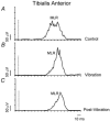
Figure 7. Averaged TA MLR evoked by a toe-down rotation under control, vibration and post-vibration conditions
A, in TA muscle an MLR can be evoked by a sudden toe-down rotation. B, during vibration, a slight and non-significant decrease of MLR with respect to control condition was observed. C, MLR was significantly depressed immediately after the vibration offset. Each trace is the average of 30 rectified and filtered EMG signals from a representative normal subject. Vertical dotted lines indicate the onset of platform movement.
Figure 3
Figure 3. Overall averages of the areas of Sol SLR and MLR under control, vibration and post-vibration conditions as percentages of maximal voluntary EMG activity (MVA), subdivided on the basis of the median values of areas of Sol background activity prior to platform rotation
The effect of vibration and post-vibration on SLR (A) and MLR (B) was evaluated separately for the trials exhibiting Sol background EMG activity lower (open columns) or higher (filled columns) than the median value. The mean areas of the background (bkg) EMGs of Sol and TA are reported in C and D, respectively. Area of the SLR was decreased during vibration and recovered towards control value during post-vibration. This behaviour was common to both background levels. MLR also decreased during vibration, but this was significant only for low background EMG activity. During post-vibration, MLR area further decreased. In this and in the following figures, * = P < 0.05, ** = P < 0.01, *** = P < 0.001.
Figure 4
Figure 4. Relationship between the areas of Sol MLR and of Sol SLR under control and vibration conditions as percentages of maximal voluntary EMG activity (MVA)
The lack of negative relationship between area of MLR and area of SLR (control condition, A) points against the notion that any decrease in the size of MLR was related to the size of the previous SLR burst. This finding is opposite to what would occur if the motoneurone pool was made refractory to a subsequent input by the SLR burst. B, under vibration conditions, the vibration-induced decrease in size of the SLR is not accompanied by an increase in size of the MLR.
Figure 5
Figure 5. Time course of the changes in area of Sol SLR (○) and MLR (•) during post-vibration as a function of the time interval between termination of vibration and onset of platform movement
The area of the SLR tended to recover immediately after vibration offset. It was still depressed, though significantly less than during vibration, at 275 ms after the end of vibration. The area of the MLR continued to decrease. Open and filled points represent the means (± s.e.m.) of the areas of Sol SLR and MLR, respectively, averaged from each subject within the corresponding 50 ms time intervals between onset of platform movement and termination of vibration. For comparison, the means of the areas of Sol SLR and MLR during control and vibration conditions are shown on the left.
Figure 8
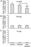
Figure 8. Overall averages of the areas of TA MLR and TA background activity and Sol background activity under control, vibration and post-vibration conditions as percentages of maximal voluntary EMG activity (MVA)
A, the effect of vibration and post-vibration on the area of TA MLR was evaluated for equal background EMG activity (bkg) of Sol (B) and TA (C). Significant depression of MLR occurred only during the post-vibration condition. This behaviour was similar to that observed in the Sol MLR during a toe-up rotation when the Sol background was greater than its median value.
Figure 9
Figure 9. Overall averages of the latency of Sol SLR and MLR and TA MLR under control (□), vibration (▪) and post-vibration (▪) conditions
The latency of Sol SLR and MLR and TA MLR was increased during vibration. This extra delay tended to vanish in the Sol responses when the vibrator was shut off. On the other hand, TA MLR showed a further delay during post-vibration.
Figure 10
Figure 10. Overall averages of the areas of Sol background, Sol SLR, Sol MLR and TA background under control (□) and 30 Hz (▪) and 90 Hz (▪) vibration conditions, as percentages of maximal voluntary EMG activity (MVA)
In spite of no difference in the area of Sol and TA background activity among the three conditions, a significant depression of SLR could be observed at 90 Hz but not at 30 Hz vibration frequency. MLR appeared to be influenced by both vibration stimuli.
Figure 11
Figure 11. Circuit diagram summarising the possible synaptic connections involved in the SLR and MLR, taking into account the effects observed during control, vibration and post-vibration conditions
For the sake of simplicity, the oligosynaptic connections fed by the group II afferents are represented by one interneurone (II-IN). White, dark grey and light grey arrows show the peripheral afferent drive and supra-spinal central drive, under the control, vibration and post-vibration conditions, respectively. Presyn-I refers to presynaptic inhibition. Note the divergent Ia input to α-motoneurones (αMN) and group II interneurones (II-IN). Further explanations are in the text.

References

    1. Allum JHJ, Büdingen HJ. Coupled stretch reflexes in ankle muscles: an evaluation of the contributions of active muscle mechanisms to human postural stability. Prog Brain Res. 1979;50:185–195. - PubMed
    1. Armitage P. Statistical Methods in Medical Research. Oxford: Blackwell Science Ltd; 1971.
    1. Ashby P, Stålberg E, Winkler T, Hunter JP. Further observations on the depression of group Ia facilitation of motoneurons by vibration in man. Exp Brain Res. 1987;69:1–6. - PubMed
    1. Bras H, Cavallari P, Jankowska E, Mccrea D. Comparison of effects of monoamines on transmission in spinal pathways from group and group II muscle afferents in the cat. Exp Brain Res. 1989;76:27–37. - PubMed
    1. Burke D, Gandevia SC, McKeon B. Monosynaptic and oligosynaptic contributions to human ankle jerk and H-reflex. J Neurophysiol. 1984;52:435–448. - PubMed

Publication types

LinkOut - more resources