. 2003 Jun 10;100(12):7325-30.
doi: 10.1073/pnas.1132092100. Epub 2003 May 30.

Neural stem cells and cholinergic neurons: regulation by immunolesion and treatment with mitogens, retinoic acid, and nerve growth factor

Affiliations

Neural stem cells and cholinergic neurons: regulation by immunolesion and treatment with mitogens, retinoic acid, and nerve growth factor

Laura Calza et al. Proc Natl Acad Sci U S A. .

Abstract

Degenerative diseases represent a severe problem because of the very limited repair capability of the nervous system. To test the potential of using stem cells in the adult central nervous system as "brain-marrow" for repair purposes, several issues need to be clarified. We are exploring the possibility of influencing, in vivo, proliferation, migration, and phenotype lineage of stem cells in the brain of adult animals with selective neural lesions by exogenous administration (alone or in combination) of hormones, cytokines, and neurotrophins. Lesion of the cholinergic system in the basal forebrain was induced in rats by the immunotoxin 192IgG-saporin. Alzet osmotic minipumps for chronic release (over a period of 14 days) of mitogens [epidermal growth factor (EGF) or basic fibroblast growth factor (bFGF)] were implanted in animals with behavioral and biochemical cholinergic defect and connected to an intracerebroventricular catheter. After 14 days of delivery, these pumps were replaced by others delivering nerve growth factor (NGF) for an additional 14 days. At the same time, retinoic acid was added to the rats' food pellets for one month. Whereas the lesion decreased proliferative activity, EGF and bFGF both increased the number of proliferating cells in the subventricular zone in lesioned and nonlesioned animals. These results are indicated by the widespread distribution of BrdUrd-positive nuclei in the forebrain, including in the cholinergic area. Performance in the water maze test was improved in these animals and choline acetyltransferase activity in the hippocampus was increased. These results suggest that pharmacological control of endogenous neural stem cells can provide an additional opportunity for brain repair. These studies also offer useful information for improving integration of transplanted cells into the mature brain.


Figures

Fig. 1.
Fig. 1.
Design of the study and composition of the experimental groups in the two sets of experiments. Time 0 refers to the day of the i.c.v. injection of 192IgG-saporin; an osmotic minipump connected to an i.c.v. catheter was implanted for EGF and bFGF administration and then replaced for NGF delivery. A diet enriched with retinoic acid (RA) was then supplied. Animals were tested for behavioral performance at the indicated time intervals and all animals were then killed for biochemical, molecular, and morphological analysis.
Fig. 3.
Fig. 3.
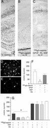
Cholinergic denervation in the hippocampus of 192IgG-lesioned animals and effects of combined treatments. Histochemical visualization of acetylcholinesterase in the hippocampal cortex of control rats (A) and 68 days after injection (B) is shown. The immunolesion induces an almost complete disappearance of esterase-reactive fibers in the hippocampus by day 68, and the deafferentation does not further worsen at 85 days (data not shown). Degeneration of cholinergic neurons in the basal forebrain is also shown (D, control, sham-operated; E, lesioned). (C) Esterase-reactive fibers were more abundant in lesioned animals after bFGF, retinoic acid (RA), and NGF administration (94 days). ChAT activity also drops in the hippocampus of lesioned animals, and combined treatment with mitogens, retinoic acid, and NGF recovers this deficit (F). In addition, the mitogens combined with retinoic acid partially recover the ChAT deficit (G), whereas single treatments do not modify ChAT activity in either lesioned or control animals. Statistical analysis was carried out by using ANOVA and Dunnett's test (P < 0.05).
Fig. 2.
Fig. 2.
The graphs report performance in the water maze task. In A, development of behavioral impairment in animals lesioned according to the paradigm used for the first set of experiments, e.g., the less severe, is illustrated by repeated tests with both visible and hidden platform. Each point represents the average of the latency over the two trials within each block. On day 20 after the injection, animals were first taught to escape to a marked, submerged platform in the water maze (visible portion of the acquisition). On the following day, the platform was in the same location but unmarked and therefore hidden from the rats' view (hidden portion of the acquisition). Three consecutive two-trial blocks of 90-sec maximum duration were run each day. Animals were then tested weekly within the hidden portion of acquisition. At 48 days after 192IgG-saporin injection, the animals showed a significant impairment in terms of latency in reaching the submerged platform (A). Repeated overall ANOVA measures between treated and untreated animals indicated a significant group effect [F(1,30) = 8.56; P < 0.005]. The lesion paradigm used in the second set of experiments produced an even more pronounced impairment in water maze performance (data not shown). Animals were then tested after combined treatments. In the first set of experiments, in which a group of lesioned animals was treated with bFGF, NGF, and retinoic acid, overall repeated ANOVA measures indicated a significant group effect [F(2,84) = 3.58; P < 0.05] at day 85 after lesion, which was evident for time spent in the platform square and not for latency (B). In the second set of experiments, in which a group of lesioned animals was treated with EGF and retinoic acid, overall repeated ANOVA measures indicated a significant group effect [F(2,80) = 3.67; P < 0.05] at day 158 after lesion (C).
Fig. 4.
Fig. 4.
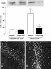
Effect of lesion and mitogen treatment on proliferative indices in the forebrain. Western blot analysis (Upper) indicates that expression of the proliferation-associated antigen Ki67 is reduced in the olfactory bulb of 192IgG-lesioned animals, and that mitogen injections expand the distribution of BrdUrd-uptaking cells in the SVZ in lesioned animals (A, control, saporin-injected; B, lesioned, 192IgG-injected + EGF).
Fig. 5.
Fig. 5.
Combined treatment in lesioned animals induces appearance of numerous small PSA-NCAM-positive cells in the basal ganglia close to the SVZ (B), whereas the same treatment in sham-operated animals is ineffective on this protein expression regulation (A). Numerous BrdUrd-uptaking cells are also observed in the basal forebrain area in lesioned animals, close to the remaining ChAT- (C) and trkA- (D) positive cells. Single cells having both nuclear labeling for BrdUrd (red) and PSA-NCAM (green) immunoreactivity were also found in the forebrain of lesioned animals receiving multiple treatments (E). To confirm the coexistence of BrdUrd and PSA-NCAM immunoreactivity, a small Inset shows the lateral projection of this double-labeling, as obtained by laser scan. No double-labeling of BrdUrd (red) with GFAP (green) was found (F).

Similar articles

Cited by

References

    1. Zhang, R. L., Zhang, Z. G., Zhang, L. & Chopp, M. (2001) Neuroscience 105, 33-41. - PubMed
    1. Jin, K., Minami, M., Lan, J. Q., Mao, X. O., Batteur, S., Simon, R. P. & Greenberg, D. A. (2001) Proc. Natl. Acad. Sci. USA 98, 4710-4715. - PMC - PubMed
    1. Arvidsson, A., Collin, T., Kirik, D., Kokaia, Z. & Lindvall, O. (2002) Nat. Med. 8, 963-970. - PubMed
    1. Yagita, Y., Kitagawa, K., Ohtsuki, T., Takasawa, K.-I., Miyata, T., Okano, H., Hori, M. & Matsumoto, M. (2001) Stroke 32, 1890-1896. - PubMed
    1. Nakatomi, H., Kuriu, T., Okabe, S., Yamamoto, S., Kawahara, N., Tamura, A., Kirino, T. & Nakafuku, M. (2002) Cell 110, 429-441. - PubMed

Publication types

MeSH terms

Grants and funding