. 2003 May 6;88(9):1411-6.
doi: 10.1038/sj.bjc.6600932.

The coxsackievirus and adenovirus receptor acts as a tumour suppressor in malignant glioma cells

Affiliations

The coxsackievirus and adenovirus receptor acts as a tumour suppressor in malignant glioma cells

M Kim et al. Br J Cancer. .

Abstract

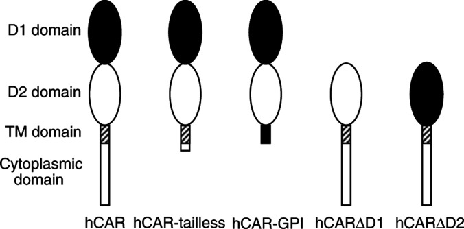
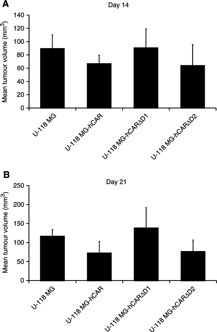
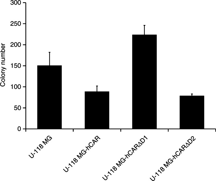
The coxsackievirus and adenovirus receptor (CAR) is a membrane glycoprotein with a cytoplasmic domain, a transmembrane domain and an extracellular region consisting of two immunoglobulin-like domains, an amino-terminal immunoglobulin variable (IgV)-related domain (D1), which is distal to the cell surface, and a proximal IgC2 domain (D2). The coxsackievirus and adenovirus receptor has been shown to exhibit tumour suppression activity in human bladder and prostate cancer cells. In the current paper, we demonstrate that CAR is a tumour suppressor in glioma cells and that the extracellular D2 domain is not required for this inhibitory effect. This finding provides a biological basis for the observation that expression of CAR is downregulated in malignant glioma cells. This suggests that strategies to redirect adenoviruses to achieve CAR-independent infection will be necessary to realise the full potential of adenoviral vectors for cancer gene therapy.


Figures

Figure 1
Figure 1
Schematic diagram of hCAR and deletion mutants. The coxsackievirus and adenovirus receptor comprises an extracellular region, a transmembrane domain (hatched rectangle) and a cytoplasmic domain (open rectangle). The extracellular region of wild-type CAR comprises an amino-terminal IgV-related domain designated D1 (solid oval), which is distal to the cell surface, and a proximal IgC2 domain designated D2 (open oval). The hCAR-tailless mutant is truncated after amino acid 260. The hCAR-GPI mutant consists of the extracellular domain of hCAR (corresponding to amino-acid residues 1–235) fused to the 37 carboxy-terminal amino acids of human DAF. The hCARΔD1 mutant lacks amino acids 21–144, while the hCARΔD2 mutant lacks amino acids 145–233 (Freimuth et al, 1999).
Figure 2
Figure 2
FACS analysis of stably transfected cells to confirm surface expression of hCAR and deletion mutants. Dark line is anti-hCAR primary monoclonal antibody RmcB plus FITC-labelled secondary antibody. Light line is FITC-labelled secondary antibody in the absence of primary antibody.
Figure 3
Figure 3
In vivo tumorigenicity assay. Tumour xenografts were established by subcutaneous injection of 5 × 10⁶ U-118 MG cells or the stably transfected derivatives into the flank of 8- to 10-week-old female athymic nu/nu nude mice (nine mice per group). Bidimensional tumour measurements were taken with calipers and the tumour volume was calculated using the simplified formula for a rotational ellipsoid: 0.5 × length × width2 (Dethlefsen et al, 1968). The mean tumour volumes±standard deviations on days 14 (A) and 21 (B) are shown. Statistical significance was achieved if P<0.05, based upon a one-way ANOVA.
Figure 4
Figure 4
Adenovirus-mediated gene transfer to U-118 MG and derivative cells. Monolayers of CAR-negative human U-118 MG malignant glioma cells were transiently transfected with pcDNA3-hCAR, pcDNA3-hCARΔD1 and pcDNA3-hCARΔD2. Forty-eight hours post-transfection, the cells were infected at a multiplicity of infection of 100 p.f.u. per cell with Ad5Luc1. After incubation for 1 h at 37°C, the vector was aspirated and the cells incubated at 37°C for 24 h. The cells were then lysed and assayed for luciferase activity, which is expressed as relative light units. Data are reported as the means±standard deviations of triplicate determinations from a representative of three independent experiments. P<0.05 was considered statistically significant.
Figure 5
Figure 5
In vivo tumorigenicity assay. Tumour xenografts were established by subcutaneous injection of 5 × 10⁶ U-118 MG cells or the stably transfected derivatives into the flank of 8- to 10-week-old female athymic nu/nu nude mice (10 mice per group). Bidimensional tumour measurements were taken with calipers and the tumour volume was calculated using the simplified formula for a rotational ellipsoid: 0.5 × length × width2 (Dethlefsen et al, 1968). The mean tumour volumes±standard deviations on days 14 (A) and 21 (B) are shown. Statistical analysis was performed by one-way ANOVA. P<0.05 was considered statistically significant.
Figure 6
Figure 6
In vitro soft agar colony-formation assay. U-118 MG cells were transfected with the various hCAR expression vectors and stable transfectants selected in the presence of 400 μg ml−1 G418. Monolayers of cells were released with Versene and dispersed into a suspension of single cells in growth medium. One thousand cells were resuspended in 2 ml of growth medium containing 0.4% low melting temperature agarose (and 400 μg ml−1 G418 in the case of the transfectants), and were overlaid in triplicate on 2 ml of solidified 0.8% low melting temperature agarose in growth medium in a 6-well dish. The dishes were incubated for 18 days at 37°C in a 5% CO2 atmosphere. Colonies larger than 50 cells were then counted. The mean colony sizes±standard deviations are shown. Statistical analysis was performed by one-way ANOVA. P<0.05 was considered statistically significant.

References

    1. Anon (1998) Use and application of adenovirus expression vectors. In Cells: A Laboratory Manual, Leinwand LA (ed) Vol. 2, pp 90.1–90.28. New York: Cold Spring Harbor Laboratory Press
    1. Barnett BG, Crews CJ, Douglas JT (2002) Targeted adenoviral vectors. Biochim Biophys Acta 1575: 1–14 - PubMed
    1. Bergelson JM, Krithivas A, Celi L, Droguett G, Horwitz MS, Wickham T, Crowell RL, Finberg RW (1998) The murine CAR homolog is a receptor for coxsackie B viruses and adenoviruses. J Virol 71: 415–419 - PMC - PubMed
    1. Blackwell JL, Miller CR, Douglas JT, Li H, Reynolds PN, Carroll WR, Peters GE, Strong TV, Curiel DT (1999) Retargeting to EGFR enhances adenovirus infection of squamous cell carcinoma of the head and neck. Arch Otolaryngol Head Neck Surg 125: 856–863 - PubMed
    1. Dethlefsen LA, Prewitt JM, Mendelsohn ML (1968) Analysis of tumor growth curves. J Natl Cancer Inst 40: 389–405 - PubMed

Publication types