

Expression of cyclin D1, D3, E, and p27 in human renal cell carcinoma analysed by tissue microarray
- PMID: 12778072
- PMCID: PMC2741051
- DOI: 10.1038/sj.bjc.6600922
Expression of cyclin D1, D3, E, and p27 in human renal cell carcinoma analysed by tissue microarray
Abstract
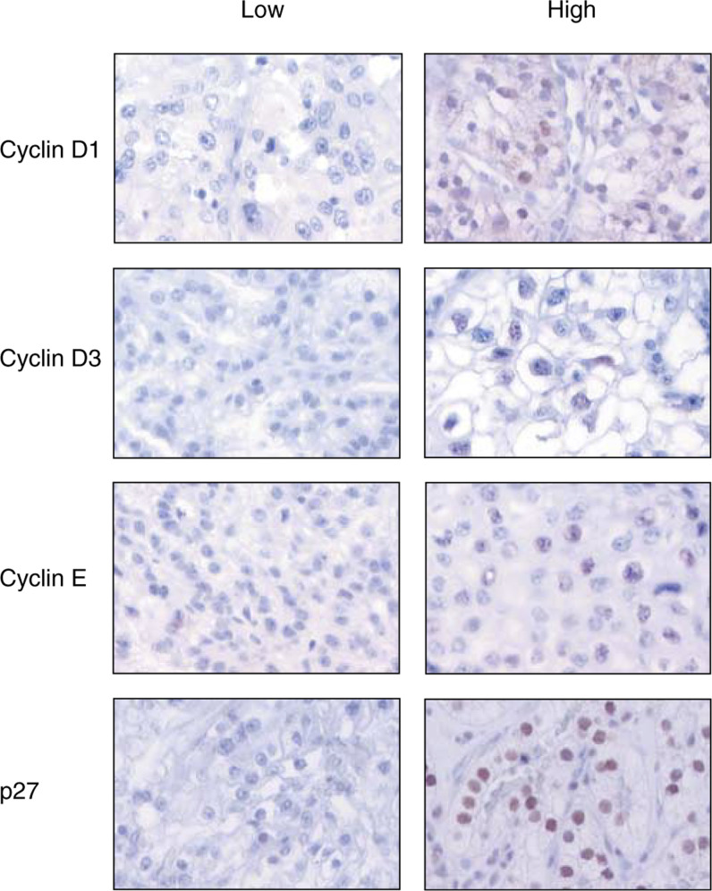
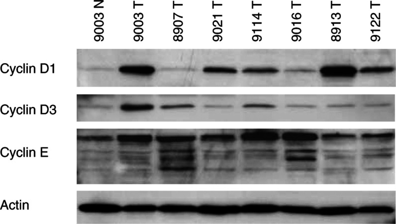
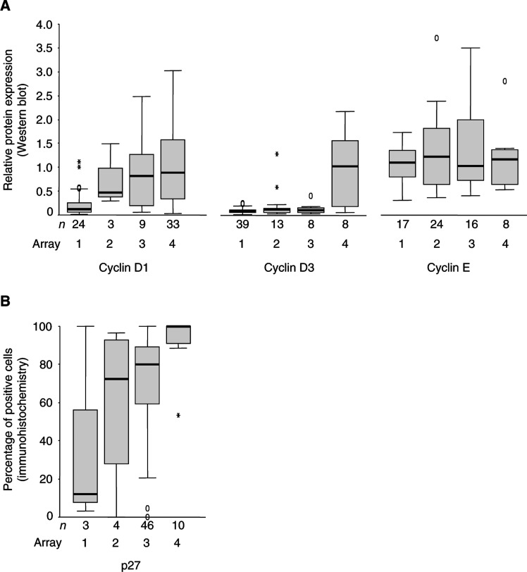
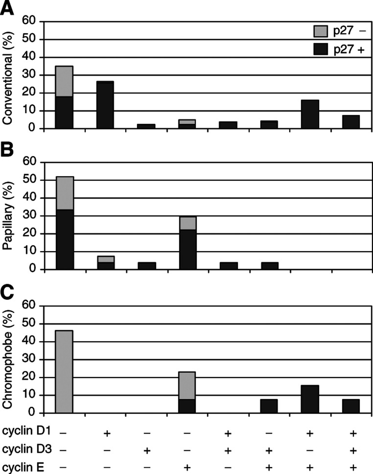
Aberrations in the G1/S transition of the cell cycle have been observed in many malignancies and seem to be critical in the transformation process. Few studies have delineated the presence of G1/S regulatory defects and their clinical relevance in renal cell carcinoma (RCC). Therefore, we have examined the protein contents of cyclin D1, D3, E, and p27 in 218 RCCs, using tissue microarray and immunohistochemistry. The results from a subset of tumours were confirmed by Western blotting and immunohistochemical staining of regular tissue sections. Interestingly, low protein contents of cyclin D1 and p27 were associated with high nuclear grade, large tumour size, and poor prognosis for patients with conventional tumours. We further observed substantial differences in the pattern of G1/S regulatory defects between the different RCC subtypes. The majority of both conventional and papillary cases expressed p27; however, chromophobe tumours generally lacked p27 staining. In addition, conventional RCCs often expressed high cyclin D1 protein levels, while papillary RCCs exhibited high cyclin E. In summary, we have shown that G1/S regulatory defects are present in RCC and are associated with clinico-pathological parameters. The pattern of cell cycle regulatory defects also differed between RCC subtypes.
Figures

References
-
- Åkervall JA, Michalides RJ, Mineta H, Balm A, Borg A, Dictor MR, Jin Y, Loftus B, Mertens F, Wennerberg JP (1997) Amplification of cyclin D1 in squamous cell carcinoma of the head and neck and the prognostic value of chromosomal abnormalities and cyclin D1 overexpression. Cancer 79: 380–389 - PubMed
-
- Bartkova J, Zemanova M, Bartek J (1996) Abundance and subcellular localisation of cyclin D3 in human tumours. Int J Cancer 65: 323–327 - PubMed
-
- Camp RL, Charette LA, Rimm DL (2000) Validation of tissue microarray technology in breast carcinoma. Lab Invest 80: 1943–1949 - PubMed
-
- Demetrick DJ, Matsumoto S, Hannon GJ, Okamoto K, Xiong Y, Zhang H, Beach DH (1995) Chromosomal mapping of the genes for the human cell cycle proteins cyclin C (CCNC), cyclin E (CCNE), p21 (CDKN1) and KAP (CDKN3). Cytogenet Cell Genet 69: 190–192 - PubMed
Publication types
MeSH terms
Substances
LinkOut - more resources
Full Text Sources
Other Literature Sources
Medical
Research Materials
