Pimonidazole binding in C6 rat brain glioma: relation with lipid droplet detection
- PMID: 12778075
- PMCID: PMC2741029
- DOI: 10.1038/sj.bjc.6600837
Pimonidazole binding in C6 rat brain glioma: relation with lipid droplet detection
Abstract
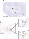
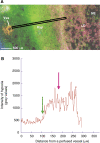
In C6 rat brain glioma, we have investigated the relation between hypoxia and the presence of lipid droplets in the cytoplasm of viable cells adjacent to necrosis. For this purpose, rats were stereotaxically implanted with C6 cells. Experiments were carried out by the end of the tumour development. A multifluorescence staining protocol combined with digital image analysis was used to quantitatively study the spatial distribution of hypoxic cells (pimonidazole), blood perfusion (Hoechst 33342), total vascular bed (collagen type IV) and lipid droplets (Red Oil) in single frozen sections. All tumours (n=6) showed necrosis, pimonidazole binding and lipid droplets. Pimonidazole binding occurred at a mean distance of 114 microm from perfused vessels mainly around necrosis. Lipid droplets were principally located in the necrotic tissue. Some smaller droplets were also observed in part of the pimonidazole-binding cells surrounding necrosis. Hence, lipid droplets appeared only in hypoxic cells adjacent to necrosis, at an approximate distance of 181 microm from perfused vessels. In conclusion, our results show that severe hypoxic cells accumulated small lipid droplets. However, a 100% colocalisation of hypoxia and lipid droplets does not exist. Thus, lipid droplets cannot be considered as a surrogate marker of hypoxia, but rather of severe, prenecrotic hypoxia.
Figures


Similar articles
-
Spatial relationship between hypoxia and the (perfused) vascular network in a human glioma xenograft: a quantitative multi-parameter analysis.Int J Radiat Oncol Biol Phys. 2000 Sep 1;48(2):571-82. doi: 10.1016/s0360-3016(00)00686-6. Int J Radiat Oncol Biol Phys. 2000. PMID: 10974478
-
Correlation between the occurrence of 1H-MRS lipid signal, necrosis and lipid droplets during C6 rat glioma development.NMR Biomed. 2003 Jun;16(4):199-212. doi: 10.1002/nbm.831. NMR Biomed. 2003. PMID: 14558118
-
Hypoxia in larynx carcinomas assessed by pimonidazole binding and the value of CA-IX and vascularity as surrogate markers of hypoxia.Eur J Cancer. 2009 Nov;45(16):2906-14. doi: 10.1016/j.ejca.2009.07.012. Epub 2009 Aug 19. Eur J Cancer. 2009. PMID: 19699082
-
Quantitative analysis of varying profiles of hypoxia in relation to functional vessels in different human glioma xenograft lines.Radiat Res. 2002 Jun;157(6):626-32. doi: 10.1667/0033-7587(2002)157[0626:qaovpo]2.0.co;2. Radiat Res. 2002. PMID: 12005540
-
Hypoxia in a human intracerebral glioma model.J Neurosurg. 2000 Sep;93(3):449-54. doi: 10.3171/jns.2000.93.3.0449. J Neurosurg. 2000. PMID: 10969943
Cited by
-
Lipid droplets and perilipins in canine osteosarcoma. Investigations on tumor tissue, 2D and 3D cell culture models.Vet Res Commun. 2022 Dec;46(4):1175-1193. doi: 10.1007/s11259-022-09975-8. Epub 2022 Jul 14. Vet Res Commun. 2022. PMID: 35834072 Free PMC article.
-
Glioblastoma metabolomics: uncovering biomarkers for diagnosis, prognosis and targeted therapy.J Exp Clin Cancer Res. 2025 Aug 7;44(1):230. doi: 10.1186/s13046-025-03497-2. J Exp Clin Cancer Res. 2025. PMID: 40775789 Free PMC article. Review.
-
Spatial profiles of markers of glycolysis, mitochondria, and proton pumps in a rat glioma suggest coordinated programming for proliferation.BMC Res Notes. 2015 Jun 2;8:207. doi: 10.1186/s13104-015-1191-z. BMC Res Notes. 2015. PMID: 26032618 Free PMC article.
-
Localized hypoxia results in spatially heterogeneous metabolic signatures in breast tumor models.Neoplasia. 2012 Aug;14(8):732-41. doi: 10.1593/neo.12858. Neoplasia. 2012. PMID: 22952426 Free PMC article.
-
Phase II Investigation of TVB-2640 (Denifanstat) with Bevacizumab in Patients with First Relapse High-Grade Astrocytoma.Clin Cancer Res. 2023 Jul 5;29(13):2419-2425. doi: 10.1158/1078-0432.CCR-22-2807. Clin Cancer Res. 2023. PMID: 37093199 Free PMC article. Clinical Trial.
References
-
- Benda P, Someda K, Messer J, Sweet WH (1971) Morphological and immunochemical studies of rat glial tumors and clonal strains propagated in culture. J Neurosurg 34: 310–323 - PubMed
-
- Callies R, Sri-Pathmanathan RM, Ferguson DYP, Brindle KM (1993) The appearance of neutral lipid signals in the 1H NMR spectra of a myeloma cell line correlates with the induced formation of cytoplasmic lipid droplets. Magn Reson Med 29: 546–550 - PubMed
-
- Chapman JD, Baer K, Lee J (1983) Characteristics of the metabolism-induced binding of misonidazole to hypoxic mammalian cells. Cancer Res 43: 1523–1528 - PubMed
-
- Durand RE, Raleigh JA (1998) Identification of nonproliferating but viable hypoxic tumor cells in vivo. Cancer Res 58: 3547–3550 - PubMed
-
- Evans S, Hahn S, Pook DR, Jenkins TW, Chalian AA, Zhang P, Stevens C, Weber R, Weinstein G, Benjamin I, Mirza N, Morgan M, Rubin S, McKenna GW, Lord EM, Koch CJ (2000) Detection of hypoxia in human squamous cell carcinoma by EF5 binding. Cancer Res 60: 2018–2024 - PubMed
Publication types
MeSH terms
Substances
LinkOut - more resources
Full Text Sources

