. 2003 Jun;132(3):485-95.
doi: 10.1046/j.1365-2249.2003.02173.x.

Release of endogenous anti-inflammatory complement regulators FHL-1 and factor H protects synovial fibroblasts during rheumatoid arthritis

Affiliations

Release of endogenous anti-inflammatory complement regulators FHL-1 and factor H protects synovial fibroblasts during rheumatoid arthritis

M A Friese et al. Clin Exp Immunol. 2003 Jun.

Abstract

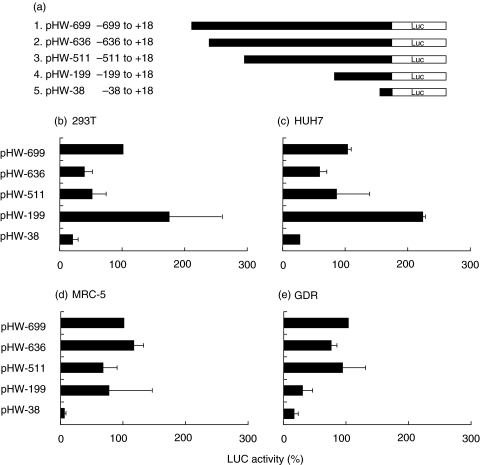
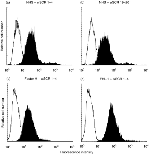
Rheumatoid arthritis is a chronic inflammatory disease of unknown aetiology predominantly affecting cells and tissues of synovial joints. Here we show that the two important complement regulators FHL-1 and factor H play a protective anti-inflammatory role in rheumatoid arthritis. Expression analyses at the mRNA- and protein level show in vitro expression and secretion of both regulators by synovial fibroblasts derived from patients with rheumatoid arthritis. Similarly the two regulators are synthesized in vivo in diseased synovial tissue, and in particular synovial lining cells express high levels of FHL-1. The anti-inflammatory role of these regulators in rheumatoid arthritis is highlighted by their induction with IFN-gamma and dexamethasone, whilst the pro-inflammatory cytokine TNF-alpha had no effect. Transient transfection experiments with various FHL-1/factor H promoter-luciferase reporter constructs into cells of distinct origin show independent cell and tissue specific promoter regulated transcription of these two regulators. The inducible expression, specifically of FHL-1 has physiological consequences. By binding directly to surfaces the released proteins protect cells from inflammatory damage and complement-mediated cell lysis. This study shows a novel protective and anti-inflammatory role of the two important complement regulators FHL-1 and factor H in rheumatoid arthritis and suggests a disease controlling role of the two proteins.


Figures

Fig. 1
Fig. 1
Expression of complement regulators in synovial fibroblasts derived from rheumatoid arthritis patients. (a) Expression of complement regulators factor H, FHL-1, CD46, CD55 and CD59 in synovial fibroblasts from rheumatoid arthritis patients was analysed at the mRNA level by RT-PCR. mRNA was isolated from synovial fibroblasts, which were either untreated (control) (lane 1), treated for 24 h with TNF-α (10 ng/ml) (lane 2), or IFN-γ (100 U/ml) (lane 3) or for 48 h with dexamethasone (0·1 µM) (lane 4). After reverse transcription sequence specific primers were used for PCR analysis. Actin-β was used as a positive control. PCR fragments were separated by agarose gel electrophoresis. (b) Analysis of FHL-1 and factor H expression at the protein level. Culture supernatants were isolated from synovial fibroblasts cultivated either in serum free medium (control) (lane 1), from cells treated for 24 h with TNF-α (10 ng/ml) (lane 2), IFN-γ (100 U/ml) (lane 3) or from cells treated for 48 h with dexamethasone (0·1 µM) (lane 4). The supernatants were separated by SDS-PAGE, transferred to nitrocellulose membranes and proteins were visualized by Western blotting using antiserum specific for factor H and FHL-1.
Fig. 2
Fig. 2
In situ expression of FHL-1 and factor H in the synovia of rheumatoid arthritis tissue. Immunohistochemistry of a section prepared from a rheumatoid arthritis patient (a,b) was compared with a respective tissue obtained from a patient with osteoarthritis, with no signs of inflammation and which was consequently used as control (c,d). Monoclonal antibody VIG8 (which recognizes an epitope located within SCR 20 of factor H) was used to locate factor H (a,c) and mAb 196X was used to detect both FHL-1 and factor H as this mAb recognizes a common epitope within SCR 1 (b,d). Staining of the synovial lining cells (especially fibroblasts and to a lesser degree macrophages) and of the interstitial spaces is seen in the RA patient (b), but not in the osteoarthritis control (d). Factor H, visualized with the specific VIG8 mAb is expressed in the interstitial spaces of the RA patient (a), while the FHL-1 and factor H recognized by mAb 196X stained the synovial lining cells, which consist particularly out of fibroblasts. No expression of the two proteins is detected in the material obtained from the osteoarthritis patients (c,d) (×40 magnification).
Fig. 3
Fig. 3
Tissue specific expression of FHL-1/factor H gene-promoter constructs. (a) Schematic diagram of the FHL-1/factor H gene promoter reporter constructs generated for reporter gene activity assays. Gene promoter activity was analysed following transient transfection of the indicated cell lines: (b) 293T kidney cells, (c) HUH7 liver cells, (d) MRC-5 lung fibroblasts and (e) GDR synovial fibroblasts. The activity was normalized to that obtained with the wild type promoter construct (pHW-699), which was set to 100%. Each column represents the mean of at least five independent experiments; the mean value and standard deviations are shown. (Results are summarized in Table 2)
Fig. 4
Fig. 4
Cell specific effect of IFN-γ on the FHL-1/factor H promoter activity. The various FHL-1/factor H promoter constructs were transiently transfected into 293T kidney cells (a), HUH7 liver cells (b), MRC-5 lung fibroblasts (c) and GDR synovial fibroblasts (d). Transfected cells were treated with interferon-γ (100 U/ml) for 24 h. The values show the increase in luciferase activity, as compared to the basal activity obtained for each construct. Each column represents the mean of at least five independent experiments; the mean value and standard deviations are shown.
Fig. 5
Fig. 5
Cell specific effect of dexamethasone on the FHL-1/factor H promoter activity. FHL-1/factor H promoter constructs were transiently transfected into 293T kidney cells (a), HUH7 liver cells (b), MRC-5 lung fibroblasts (c) and GDR synovial fibroblasts (d). Transfected cells were treated with dexamethasone (0·1 µM) for 24 h. The values show the increase in luciferase activity, as compared to the basal activity obtained for each construct. Each column represents the mean of at least five independent experiments; the mean value and standard deviations are shown.
Fig. 6
Fig. 6
Effect of TNF-α on the FHL-1/factor H promoter activity in synovial fibroblasts. FHL-1/factor H promoter constructs were transiently transfected into GDR synovial fibroblasts which were further treated with TNF-α (10 ng/ml) for 24 h. The values represent the increase in luciferase activity compared with the basal activity of each construct (1×). Each column represents the mean of at least five independent experiments; the mean value and standard deviations are shown.
Fig. 7
Fig. 7
Binding of FHL-1 and factor H to the surface of synovial fibroblasts. Binding of FHL-1 and factor H to synovial fibroblasts was analysed by flow cytometry. Cells cultured in serum free medium were used directly (control) or incubated with 25% NHS (a,b), purified complement factor H (500 µg/ml) (c) or purified recombinant FHL-1 (50 µg/ml) (d). After washing cells were stained with specific polyclonal antiserum directed against SCR 1–4 (a,c,d) or SCR 19–20 (b), followed by FITC-labelled swine anti-rabbit antiserum (▪). Irrelevant mouse IgG was used as a primary antibody for control staining (□).
Fig. 8
Fig. 8
The effect of FHL-1 and factor H neutralization on complement-mediated killing of synovial fibroblasts cells. 51Cr labelled cells were incubated with varying dilutions of anti-SCR 1–4 (•) or anti-SCR 19–20 (○) antibodies and in 25% NHS as the complement source. Cell lysis was determined after 30 min incubation by 51Cr release. The results represent means ± SD of duplicate experiments.

References

    1. Müller-Eberhard HJ, Schreiber RD. Molecular biology and chemistry of the alternative pathway of complement. Adv Immunol. 1980;29:1–53. - PubMed
    1. Walport MJ. Complement. N Engl J Med. 2001;344:1140–4. - PubMed
    1. Rus HG, Niculescu FI, Shin ML. Role of the C5b-9 complement complex in cell cycle and apoptosis. Immunol Rev. 2001;180:49–55. - PubMed
    1. Kraus S, Seger R, Fishelson Z. Involvement of the ERK mitogen-activated protein kinase in cell resistance to complement-mediated lysis. Clin Exp Immunol. 2001;123:366–74. - PMC - PubMed
    1. Zipfel PF, Skerka C. FHL-1: a human complement and immune regulator with cell-adhesive function. Immunol Today. 1999;20:135–40. - PubMed

Publication types

MeSH terms