

Histopathologic features of Mycobacterium ulcerans infection
- PMID: 12780997
- PMCID: PMC3000137
- DOI: 10.3201/eid0906.020485
Histopathologic features of Mycobacterium ulcerans infection
Abstract
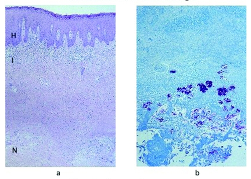
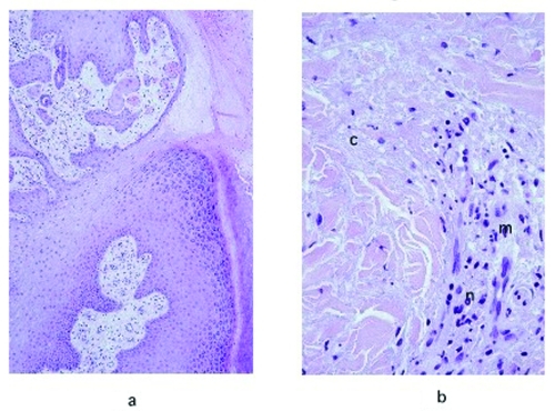
Because of the emergence of Buruli ulcer disease, the World Health Organization launched a Global Buruli Ulcer Initiative in 1998. This indolent skin infection is caused by Mycobacterium ulcerans. During a study of risk factors for the disease in Ghana, adequate excisional skin-biopsy specimens were obtained from 124 clinically suspicious lesions. Buruli ulcer disease was diagnosed in 78 lesions since acid-fast bacilli (AFB) were found by histopathologic examination. Lesions with other diagnoses included filariasis (3 cases), zygomycosis (2 cases), ulcerative squamous cell carcinomas (2 cases), keratin cyst (1 case), and lymph node (1 case). Thirty-seven specimens that did not show AFB were considered suspected Buruli ulcer disease cases. Necrosis of subcutaneous tissues and dermal collagen were found more frequently in AFB-positive specimens compared with specimens from suspected case-patients (p<0.001). Defining histologic criteria for a diagnosis of Buruli ulcer disease is of clinical and public health importance since it would allow earlier treatment, leading to less deforming sequelae.
Figures
References
-
- Carey MJ, Connor DH. Buruli ulcer—infection with Mycobacterium ulcerans. In: Connor DH, Chandler FW, Schwartz DA, Manz HJ, Lack EE, editors. Pathology of infectious diseases. Stamford (CT): Appleton & Lange; 1997. p. 453–9.
-
- King CH, Ashford DA, Dobos KM, Spotts Whitney EA, Raghunathan PL, Guarner J, et al. Mycobacterium ulcerans infection and Buruli ulcer disease: emergence of a public health dilemma. In: Scheld WM, Craig WA, Hughes JM, editors. Emerging infections. Washington: American Society of Microbiology Press; 2001;9:137–52.
-
- Asiedu K, Scherpbier R, Raviglione M. Buruli ulcer, Mycobacterium ulcerans infection. Geneva: World Health Organization; 2000.
Publication types
MeSH terms
Substances
LinkOut - more resources
Full Text Sources
Medical