. 2003 Jun 1;17(11):1328-33.
doi: 10.1101/gad.1077103.

TRF1 is degraded by ubiquitin-mediated proteolysis after release from telomeres

Affiliations

TRF1 is degraded by ubiquitin-mediated proteolysis after release from telomeres

William Chang et al. Genes Dev. .

Abstract

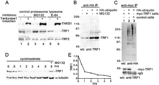
Mammalian telomeres are coated by the sequence-specific, DNA-binding protein, TRF1, a negative regulator of telomere length. Previous results showed that ADP-ribosylation of TRF1 by tankyrase 1 released TRF1 from telomeres and promoted telomere elongation. We now show that loss of TRF1 from telomeres results in ubiquitination and degradation of TRF1 by the proteasome and that degradation is required to keep TRF1 off telomeres. Ubiquitination of TRF1 is regulated by its telomere-binding status; only the telomere-unbound form of TRF1 is ubiquitinated. Our findings suggest a novel mechanism of sequential post translational modification of TRF1 (ADP-ribosylation and ubiquitination) for regulating access of telomerase to telomeres.


Figures

Figure 1.
Figure 1.
Tankyrase 1-induced telomere elongation is telomerase-dependent. (A–C) Generation of a stable WI38 cell line expressing telomerase. WI38 cells at PD 4 expressing vector control (V) or telomerase (TERT) were analyzed for telomerase expression by immunoblotting whole-cell extracts with anti-TERT 374 antibody (A), or for telomerase activity by TRAP (telomere repeat amplification protocol; B) or for telomere length by Southern blot analysis (C). (D–F) Analysis of stable WI38 cells expressing telomerase (WI38-TERT) and tankyrase 1 alleles. WI38-TERT cells expressing vector control (V), FN-tankyrase1.WT (WT), or FN-tankyrase1.HE/A (HE/A) were analyzed at PD 9 by immunoblotting whole-cell extracts with the antibodies anti-tankyrase 1 376 (TNKS1), anti-poly(ADP-ribose) (PAR), anti-telomerase (TERT), or anti-TRF1 415 (D); or at PD 11 for telomerase activity by TRAP (E); or at the indicated PDs for telomere length by Southern blot analysis (F).
Figure 2.
Figure 2.
TRF1 degradation by the ubiquitin-proteasome pathway. (A) TRF1 is degraded by the proteasome in vivo. Immunoblot analysis of cell extracts from FN30 cells grown in the absence (-) or presence (+) of tankyrase 1 induction. Prior to harvesting, cells were treated without (-) or with (+) proteasome inhibitor (12.5 μM MG132) or with (+) lysosomal inhibitor (12.5 μM E-64) for 10 h. (B,C) TRF1 is ubiquitinated in vivo. (B) HelaI.2.11 cells were transfected with (+) or without (-) a plasmid expressing HA-ubiquitin for 24 h. Four hours prior to harvest, cells were treated with (+) or without (-) 12.5 μM MG132, lysed, and incubated with anti-HA affinity matrix. (C) HTC75 cells expressing myc-TRF1 or a vector control were transfected with (+) or without (-) HA-ubiquitin for 24 h, lysed and incubated with anti-myc affinity matrix. (A–C) Proteins were fractionated on SDS-PAGE and analyzed by Western blotting (wb) with anti-tankyrase 1 376 (TNKS1), anti-TRF1 415, anti-TRF2, or anti-HA antibodies. (B,C) Black dot indicates ubiquitinated TRF1. (D) TRF1 has a short half-life. HT1080 cells were incubated with 100 μg/mL cycloheximide for 0–5 h or for 5 h with 100 μg/mL cycloheximide and 12.5 μM MG132 (+). Cells were harvested, lysed, and cell extracts were fractionated by SDS-PAGE and analyzed by immunoblotting with anti-TRF1 415 or anti-α-tubulin antibodies. (E) Graphical representation of relative TRF1 levels normalized against the α-tubulin loading control using Image Quant version 1.2. The graph represents an average of four experiments. Bars represent the average ± standard error calculated from four independent experiments.
Figure 3.
Figure 3.
In vitro ubiquitination of TRF1 is not dependent on ADP-ribosylation. (A-B) TRF1 is ubiquitinated in vitro. 35S-labeled TRF1 was generated by coupled in vitro transcription/translation and then diluted into a ubiquitination reaction mix containing reticulocyte lysate supplemented with methyl ubiquitin, ubiquitin aldehyde, and an energy source [retic(+)] without (-) or with (+) exogenous ubiquitin. (C) TRF1 interaction with tankyrase 1 is not required for ubiquitination. 35S-labeled TRF1 (C) or Δacidic TRF1 (Δ) was incubated without (-) or with (+) retic(+) ubiquitination reaction mix. (D) TRF1 is ubiquitinated in the presence of the PARP inhibitor 3AB. 35S-labeled in vitro translated TRF1 generated in the presence of 0, 1, or 10 mM 3AB was incubated without (-) or with (+) retic(+) ubiquitination reaction mix containing 0, 1, or 10 mM 3AB. Following incubations proteins were fractionated on SDS-PAGE and visualized by fluorography (A,C,D) or analyzed by immunoblotting (B) with anti-TRF1 415 antibody.
Figure 4.
Figure 4.
Telomeric TRF1 is protected from ubiquitination. (A) Ubiquitination of TRF1 is specifically inhibited by binding to double-stranded telomeric TTAGGG repeats. In vitro ubiquitination reactions containing 35S-labeled TRF1 were carried out in the absence (-) or presence of double-stranded (DS; 0, 0.01, 0.05, 0.25, or 1 μg TTAGGG, or 0.25 or 1 μg TTAGGC) repeat DNA or single-stranded [SS; 1 μg TTAGGG (G) or AATCCC (C)] repeat DNA. (B) Ubiquitination of a TRF1 DNA-binding mutant is not inhibited by telomeric DNA. 35S-labeled TRF1.RV was incubated without (-) or with [+; retic(+)] ubiquitination reaction mix in the absence (-) or presence of double-stranded (0.25 or 1 μg TTAGGG) repeat DNA. (C) Ubiquitination of TRF1 lacking the myb domain is greatly reduced. 35S-labeled TRF1 (C) or ΔmybTRF1 (Δmyb) was incubated without (-) or with (+) retic(+) ubiquitination reaction mix. (A–C) Following incubations, proteins were fractionated on SDS-PAGE and visualized by fluorography. (D,E) Proteasome inhibition attenuates tankyrase 1-induced loss of telomeric TRF1. HelaI.2.11 cells were transiently transfected with FN-tankyrase1.WT for 24 h. Eight hours prior to harvest, cells were incubated without (-; D) or with (+; E) 25 μM MG132 and analyzed by indirect immunofluorescence. Paraformaldehyde-fixed cells were stained with anti-FLAG (green) or anti-TRF1 415 (red) antibodies. DAPI staining of DNA is shown in blue. Bar, 5 μm.
Figure 5.
Figure 5.
Model: sequential posttranslational modification of TRF1 regulates access of telomerase to telomeres. TRF1-bound telomeres are inaccessible to telomerase. Tankyrase 1 ADP-ribosylates TRF1, removing it from telomeres. Once TRF1 is off telomeres, it is ubiquitinated and degraded by the proteasome. TRF1-free telomeres are elongated by telomerase and then reassembled into a telomerase-inaccessible state using newly synthesized TRF1.

References

    1. Ancelin K., Brunori, M., Bauwens, S., Koering, C.E., Brun, C., Ricoul, M., Pommier, J.P., Sabatier, L., and Gilson, E. 2002. Targeting assay to study the cis functions of human telomeric proteins: Evidence for inhibition of telomerase by TRF1 and for activation of telomere degradation by TRF2. Mol. Cell. Biol. 22: 3474–3487. - PMC - PubMed
    1. Bilaud T., Brun, C., Ancelin, K., Koering, C.E., Laroche, T., and Gilson, E. 1997. Telomeric localization of TRF2, a novel human telobox protein. Nat. Genet. 17: 236–239. - PubMed
    1. Broccoli D., Smogorzewska, A., Chong, L., and de Lange, T. 1997. Human telomeres contain two distinct Myb-related proteins, TRF1 and TRF2. Nat. Genet. 17: 231–235. - PubMed
    1. Cenci G., Rawson, R.B., Belloni, G., Castrillon, D.H., Tudor, M., Petrucci, R., Goldberg, M.L., Wasserman, S.A., and Gatti, M. 1997. UbcD1, a Drosophila ubiquitin-conjugating enzyme required for proper telomere behavior. Genes & Dev. 11: 863–875. - PubMed
    1. Chong L., van Steensel, B., Broccoli, D., Erdjument-Bromage, H., Hanish, J., Tempst, P., and de Lange, T. 1995. A human telomeric protein. Science 270: 1663–1667. - PubMed

Publication types

MeSH terms

LinkOut - more resources