. 2003 Jun 1;17(11):1415-28.
doi: 10.1101/gad.1056603.

Yeast enhancer of polycomb defines global Esa1-dependent acetylation of chromatin

Affiliations

Yeast enhancer of polycomb defines global Esa1-dependent acetylation of chromatin

Alexandre A Boudreault et al. Genes Dev. .

Abstract

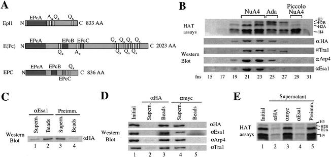
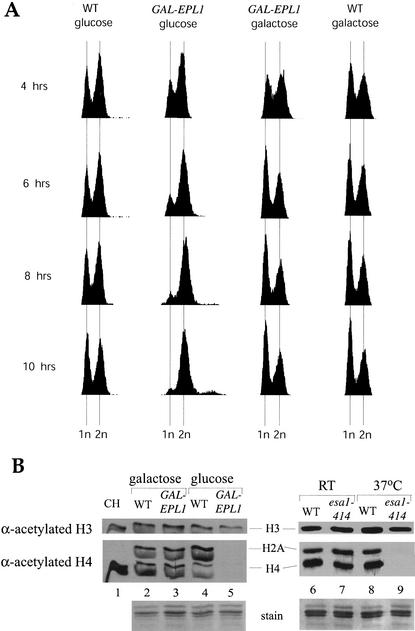
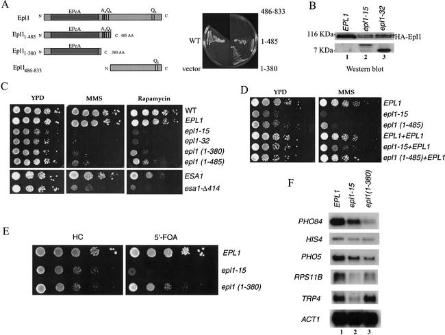
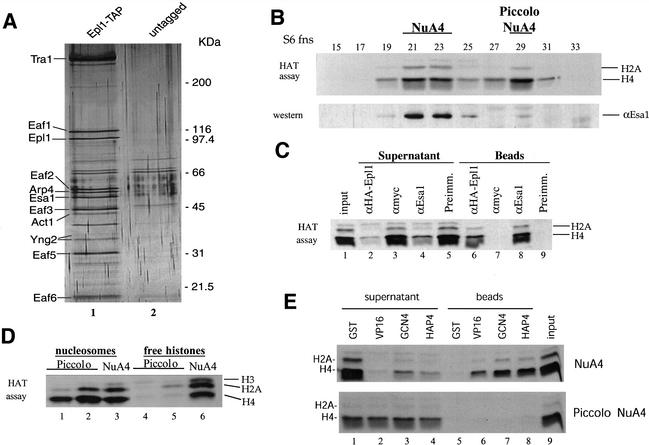
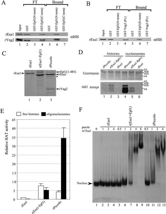
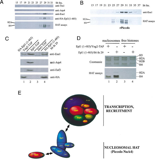
Drosophila Enhancer of Polycomb, E(Pc), is a suppressor of position-effect variegation and an enhancer of both Polycomb and trithorax mutations. A homologous yeast protein, Epl1, is a subunit of the NuA4 histone acetyltransferase complex. Epl1 depletion causes cells to accumulate in G2/M and global loss of acetylated histones H4 and H2A. In relation to the Drosophila protein, mutation of Epl1 suppresses gene silencing by telomere position effect. Epl1 protein is found in the NuA4 complex and a novel highly active smaller complex named Piccolo NuA4 (picNuA4). The picNuA4 complex contains Esa1, Epl1, and Yng2 as subunits and strongly prefers chromatin over free histones as substrate. Epl1 conserved N-terminal domain bridges Esa1 and Yng2 together, stimulating Esa1 catalytic activity and enabling acetylation of chromatin substrates. A recombinant picNuA4 complex shows characteristics similar to the native complex, including strong chromatin preference. Cells expressing only the N-terminal half of Epl1 lack NuA4 HAT activity, but possess picNuA4 complex and activity. These results indicate that the essential aspect of Esa1 and Epl1 resides in picNuA4 function. We propose that picNuA4 represents a nontargeted histone H4/H2A acetyltransferase activity responsible for global acetylation, whereas the NuA4 complex is recruited to specific genomic loci to perturb locally the dynamic acetylation/deacetylation equilibrium.


Figures

Figure 1.
Figure 1.
Epl1 is a stable stoichiometric subunit of the NuA4 complex. (A) Diagram of homology regions of yeast Epl1 compared with Drosophila E(Pc) and human EPC. (B) Epl1 coelutes with NuA4 components and H4/H2A acetyltransferase activity. Whole cell extract from a HA-Epl1-expressing strain was fractionated over nickel-agarose, followed by a MonoQ column. Peak NuA4 fractions were pooled and loaded on a Superose 6 gel-filtration column. Fractions were tested for HAT activity and by Western blotting with the indicated antisera. (C) Epl1 is a stable Esa1-associated factor. Equal amounts of Superose 6 fraction 21 described in B were incubated with preimmune or anti-Esa1 beads. The majority of HA-Epl1 coimmunoprecipitates with Esa1. (D) HA-Epl1 coimmunoprecipitates Esa1, Arp4, Tra1, and NuA4 HAT activity, showing that Epl1 is a stable stoichiometric subunit of NuA4. The Superose 6 fraction was incubated with anti-HA or anti-Myc beads. After washes, equivalent amounts of initial samples, beads, and supernatant were analyzed by Western blotting with anti-HA, anti-Esa1, anti-Arp4, and anti-Tra1 as indicated. (E) Immunoprecipitation of HA-Epl1 fully depletes NuA4 HAT activity from the supernatant. Immunoprecipitations were performed as in C and D, and initial samples and supernatants were tested for NuA4 HAT activity.
Figure 2.
Figure 2.
Epl1 expression is essential for cell cycle progression and global histone H4 and H2A acetylation in chromatin. (A) Epl1 is required for progression through mitosis. FACS analysis of cells expressing Epl1 under control of the GAL1 promoter and the isogenic wild type for the indicated times in either galactose or glucose media. When Epl1 expression is repressed in glucose, cells accumulate in G2/M. (B) Histones were purified from wild-type or GAL1-EPL1-expressing cells after 12 h in galactose or glucose media and run on an 18% SDS-PAGE. Histones purified from wild-type and esa1 ts mutant cells grown at room temperature or at 37°C for 4 h were also examined for comparison. Gels were blotted and screened with the indicated antibodies.
Figure 3.
Figure 3.
Epl1 mutations affect growth, sensitivity to DNA damage, target gene transcription, and telomeric silencing. (A) Schematic presentation of Epl1 truncations that were tested in growth complementation assay. Only the conserved EPc domain of Epl1 is required for cell viability. EPL1-deleted haploid strains expressing wild-type Epl1 from a URA3 vector and the indicated Epl1 truncations from a low-copy LEU2 vector were selected on 5′-FOA plates to evict the URA3 plasmid. (B) epl1 mutant clones obtained in a screen produce low amounts of full-length Epl1 protein. Western analysis of extracts from epl1-15 and epl1-32 mutants show that both strains produce a very short truncated protein, as expected based on the mutation, but also low amounts of full-length Epl1. The asterisk indicates a nonspecific band detected by anti-HA. (C) Cells expressing Epl1 C-terminal truncation and dosage mutants exhibit slow growth and are highly sensitive to MMS and rapamycin. Here, 10-fold serial dilutions of the EPL1, epl1-15, epl1-32, epl1 (1–380), and epl1 (1–485) strains were spotted on YPD, YPD + 0.03% MMS, or YPD + 25 nM rapamycin plates and incubated at 30°C for 2 d (YPD) and 4 d (MMS and rapamycin). esa1-Δ414 mutant cells were also tested and show the same sensitivity to the drugs. (D) The dominance/recessivity aspect of the epl1 mutants was tested by coexpression of the wild-type protein in the mutant cells. Spot dilution analyses as in C were performed and show that wild-type Epl1 rescues growth in the mutants, indicating that they are not dominant. (E) The conserved EPc domain of Epl1 is important for telomeric silencing. The 10-fold serial dilutions of the strains QY129 (EPL1), QY130 (epl1-15), and QY131 [epl1 (1–380)] containing a URA3 gene located next to the telomere region of Chromosome VII were spotted on Hartwell's complete (HC) or HC + 5′-FOA plates (to reflect expression of URA3) and incubated at 30°C for 2 and 3 d, respectively. (F) Epl1 is required for normal expression of specific genes in vivo. RNA was extracted from the indicated strains and analyzed by Northern blot using PHO84, HIS4, PHO5, RPS11B, TRP4, and ACT1 probes.
Figure 4.
Figure 4.
Epl1 is also present with Esa1 in Piccolo NuA4, a small highly active, chromatin-preferring, nontargeted histone H4/H2A acetyltransferase complex. (A) Soluble Epl1 is exclusively associated with NuA4 components in the cell. A yeast strain expressing Epl1 with a TAP (tandem affinity purification) cassette at its C terminus was produced by homologous recombination. Purified TAP-Epl1 and its associated proteins were run on SDS-PAGE and silver stained. Known NuA4 components are labeled on the left based on molecular weight and Western and mass spectrometric analysis. Fractionation from a control untagged strain is also presented to indicate nonspecific bands. (B) Identification of Piccolo NuA4 as a new histone acetyltransferase complex. Whole cell yeast extract purified over nickel-agarose followed by MonoQ was subsequently passed over a Superose 6 gel filtration column. Fractions were assayed for HAT activity (top panel) and Western blotted for the presence of Esa1 (bottom panel). An additional nucleosomal H4/H2A acetyltransferase activity is apparent in fraction 29. The 300-kD complex (Piccolo NuA4) has a similar level of HAT activity as NuA4 (fractions 21–23; ∼10,000 cpm/μL). Note that a low amount of Esa1 is also detected by Western blot in fraction 29. (C) Esa1 and Epl1 are stable components of Piccolo NuA4 as demonstrated by coimmunoprecipitation of picNuA4 HAT activity with Esa1 and HA-Epl1. Equal amounts of the Superose 6 fraction 29 obtained from an HA-Epl1-expressing strain were incubated with anti-HA, anti-Esa1, anti-Myc, and preimmune serum beads. After washes, equivalent amounts of input sample, supernatant, and beads were used in nucleosomal HAT assays. (D) NuA4 and picNuA4 complexes differ in their ability to acetylate histones. Complexes were immunopurified (with anti-Esa1 beads) from Superose 6 peak fractions of NuA4 and two different picNuA4 preps and assayed for HAT activity on oligonucleosomes or free histones. Although NuA4 acetylates free histones and nucleosomal histones at relatively similar levels (lanes 3,6), picNuA4 has a striking preference for chromatin (lanes 1,2 vs. 4,5). (E) Unlike NuA4, picNuA4 does not interact with the acidic transcription activation domains of VP16, Gcn4, and Hap4 in vitro. NuA4 or picNuA4 Superose fractions were incubated with GST-VP16, GST-Gcn4, GST-Hap4, or control GST beads. Supernatant was removed, beads were washed, and equivalent amounts were assayed for nucleosomal HAT activity.
Figure 5.
Figure 5.
The “Enhancer of Polycomb” domain of Epl1 bridges Esa1 and Yng2 to enable nucleosomal acetyltransferase activity. (A,B) GST pull-downs using a near stoichiometric ratio of the indicated recombinant proteins were carried out and assayed by Western blotting. As shown using αHIS Western blots, both rEsa1p and rYng2p are pulled down using GST-Epl1 (N-term) but not using the C terminus of the same protein, and GST-Yng2 (FL) does not associate with rEsa1. (C) Coomassie staining of the purified recombinant proteins and bacterially coexpressed complexes used for HAT and mobility shift assays. The slight size difference of rEsa1 in lane 1 versus lanes 2 and 3 is caused by different cloning strategies. The band marked by an asterisk is a degradation product of rEpl1. The loaded amount of proteins as standardized to rEsa1 is 4, 8, and 8 pmole (lanes 1,2,3, respectively). (D) Relative HAT activity of rEsa1 alone versus polycistronic recombinant complexes. HAT assays with the recombinant proteins shown in C were carried out with free histones or nucleosomes. rEsa1 was kept constant in all assays. (E) Quantification of HAT assays done on free (open box) or nucleosomal (black box) histones. Data are representative of two different experiments done in duplicate. (F) Mobility shift assay with mononucleosomes shows the formation of ternary complexes: Nucleosome binding is detected with r(Esa1–Epl1) (lanes 59) and rPiccolo (lanes 1013), whereas rEsa1 shows no interaction (lanes 24).
Figure 6.
Figure 6.
Essential functions of Esa1 and Epl1 through their presence in the Piccolo NuA4 complex. (A) The C-terminal portion of Epl1 is important to maintain integrity of the NuA4 complex. Extract from an epl1 deletion strain covered by a HA-Epl1 (1–485)-expressing plasmid, was purified over nickel-agarose, followed by MonoQ column fractionation. MonoQ peak fractions were pooled and loaded on a Superose 6 gel filtration column. Fractions were tested for HAT activity and analyzed by Western blotting with the indicated antisera. (B) Recombinant Piccolo NuA4 from the Source S peak fraction was loaded on a Superose 6 filtration column; fractions were analyzed as in A. (C) Tandem affinity purification of Yng2 from epl1 (1–485) cells brings only picNuA4 components. Western analysis of TAP-purified material obtained with YNG2–TAP/HA–EPL1 (1–485), EPL1–TAP (NuA4 control), and DOT1–TAP (control) cells. NuA4-specific components are only detected in EPL1–TAP cells. Superose 6 fraction 29 from A is also shown for comparison. (D) Affinity- and conventionally purified picNuA4-like complexes from A and C were tested in a HAT assay on oligonucleosomes and free histones. Both complexes show strong preference for chromatin substrates (cf. lanes 1,2 and 3,4). (E) Model of Piccolo NuA4 anchoring itself to the rest of the NuA4 complex through the Epl1 C-terminal region. Eaf3 is depicted separately because of a previously reported direct interaction with Esa1 (Eisen et al. 2001). Subunit names are indicated, and protein domains are italicized. EPC, conserved “Enhancer of Polycomb” domain; PHD, PHD finger domain; HAT, histone acetyltransferase domain; CHD, chromo domain; AID, activator interaction domain; PI-3K, phospho-inositol-3 kinase family domain.

References

    1. Adams A., Gottschling, D.E., Kaiser, C.A., and Stearns, T. 1997. Methods in yeast genetics. Cold Spring Harbor Laboratory Press, Cold Spring Harbor, NY.
    1. Allard S., Utley, R.T., Savard, J., Clarke, A., Grant, P., Brandl, C.J., Pillus, L., Workman, J.L., and Côté, J. 1999. NuA4, an essential transcription adaptor/histone H4 acetyltransferase complex containing Esa1p and the ATM-related cofactor Tra1p. EMBO J. 18: 5108–5119. - PMC - PubMed
    1. Balasubramanian R., Pray-Grant, M.G., Selleck, W., Grant, P.A., and Tan, S. 2002. Role of the Ada2 and Ada3 transcriptional coactivators in histone acetylation. J. Biol. Chem. 277: 7989–7995. - PubMed
    1. Bird A.W., Yu, D.Y., Pray-Grant, M.G., Qiu, Q., Harmon, K.E., Megee, P.C., Grant, P.A., Smith, M.M., and Christman, M.F. 2002. Acetylation of histone H4 by Esa1 is required for DNA double-strand break repair. Nature 419: 411–415. - PubMed
    1. Brown C.E., Howe, L., Sousa, K., Alley, S.C., Carrozza, M.J., Tan, S., and Workman, J.L. 2001. Recruitment of HAT complexes by direct activator interactions with the ATM-related Tra1 subunit. Science 292: 2333–2337. - PubMed

Publication types