. 2003 Jun;111(11):1673-81.
doi: 10.1172/JCI17098.

In vivo administration of a lentiviral vaccine targets DCs and induces efficient CD8(+) T cell responses

Affiliations

In vivo administration of a lentiviral vaccine targets DCs and induces efficient CD8(+) T cell responses

Christoph Esslinger et al. J Clin Invest. 2003 Jun.

Abstract

The present study evaluates the potential of third-generation lentivirus vectors with respect to their use as in vivo-administered T cell vaccines. We demonstrate that lentivector injection into the footpad of mice transduces DCs that appear in the draining lymph node and in the spleen. In addition, a lentivector vaccine bearing a T cell antigen induced very strong systemic antigen-specific cytotoxic T lymphocyte (CTL) responses in mice. Comparative vaccination performed in two different antigen models demonstrated that in vivo administration of lentivector was superior to transfer of transduced DCs or peptide/adjuvant vaccination in terms of both amplitude and longevity of the CTL response. Our data suggest that a decisive factor for efficient T cell priming by lentivector might be the targeting of DCs in situ and their subsequent migration to secondary lymphoid organs. The combination of performance, ease of application, and absence of pre-existing immunity in humans make lentivector-based vaccines an attractive candidate for cancer immunotherapy.


Figures

Figure 1
Figure 1
Tissue distribution of lentivector obtained after in vivo administration into footpads of mice. (a) Detection of integrated lentivector at the site of injection (foot), the draining lymph node, and the spleen but not in the mandibular lymph node and the tip of the tail. Vector integration was detected by amplification of vector-derived GFP sequence with specific primers (arrows) using seminested PCR. dLN, draining lymph node; spl, spleen; man, mandibular lymph node. (b) Histological analysis of GFP expression after administration of lentivector into one hind footpad. The upper panel shows GFP fluorescence of frozen sections of popliteal lymph nodes 2.5 days after injection of CMV-GFP lentivector (lvGFP) or PBS (nontreated) into the footpad. The contralateral popliteal lymph node (contra) of a treated mouse is also shown. Magnification, ×100. The lower panel shows double immunofluorescence analysis of draining lymph nodes and spleen 2.5 days after injection of CMV-GFP lentivector using anti-GFP antibodies (green) counterstained with anti-CD11c, anti-CD11b, and anti-B220 antibodies (red). Magnification, ×400. The use of a CMV-GFP lentivector for immunofluorescence was necessary, because with the bicistronic vaccine constructs (antigen-IRES-GFP), GFP fluorescence was too low to be picked up in histology. For PCR analysis of the GFP sequence, the actual vaccine vector was used.
Figure 2
Figure 2
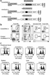
Direct administration of lentivector results in the induction of Cw3 CTL responses. (a) Schematic representation of the constructs used to generate lentiviral vectors. (b) Phenotypic characterization of peak response (day 9). PBLs were analyzed for the cell surface markers CD8, CD62L (downregulated upon activation), and TCR Vβ10. The T cell receptor specificity was examined using H-2Kd/Cw3 tetramers in combination with Vβ10 staining. The proportion of Cw3-specific cells among total PBLs obtained after direct in vivo administration of lentivector is depicted. As a control, an irrelevant (miniMelan-A) lentivector was administered. This is followed by a detailed analysis of the immune response, featuring the proportion of activated CD8+ cells, the proportion of Cw3-specific cells in the activated compartment, and the proportion of Cw3-specific cells among total CD8+ cells. (c) CTL assay showing the in vivo elimination of target cells transferred to vaccinated mice. Syngeneic splenocytes, pulsed with Cw3 antigenic peptide (RYLKNGKETL) and labeled with CFSE at high concentration, were transferred to vaccinated mice 1 day before peak response along with the same number of nonpulsed splenocytes labeled with CFSE at a lower concentration. Twelve hours later, the disappearance of peptide-pulsed cells was determined by FACS analysis in PBL, spleen, and liver. By a comparison of the ratio of pulsed to nonpulsed cells, the percentage of specific killing was calculated. Control lv, lentivector expressing an irrelevant CTL epitope; miniMelan-A lv, ELA26–35 minigene lentivector.
Figure 3
Figure 3
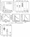
Comparative analysis of the CD8+ T cell response as induced by lentivector administration, DC-based vaccination, and peptide/CpG/adjuvant vaccination. (a) Comparative vaccination with transgenic DCs, transduced DCs, and direct in vivo administration of lentivector. Mice were vaccinated and PBLs were analyzed by FACS analysis as described. Evolution of the Cw3-specific population over time is shown. All graphs show individual mice that were representative of at least seven mice used for each condition in at least three independent experiments. (b) Proportion of Cw3-specific cells in the CD8+ compartment as determined at peak response with all vaccination conditions plus preimmune levels (dashed lines and x = average, n = number of mice tested). (c) Comparative vaccination with Cw3 minigene lentivector and Cw3 peptide/CpG/IFA vaccination. Shown are time courses of the proportion of activated CD8+ cells and the proportion of Cw3-specific cells within the activated population followed by the proportion of Cw3-specific cells in the CD8+ population. (d) Proportion of Cw3-specific cells in the CD8+ compartment as determined at peak response (day 9 for lentivector vaccination and day 7 for peptide-adjuvant vaccination) of all mice tested (dashed lines and x = average, n = number of mice tested).
Figure 4
Figure 4
Induction of a Melan-A–specific CD8+ T cell response in HLA-A*0201/Kb transgenic mice vaccinated by administration of lentivector encoding the Melan-A CTL epitope ELA26–35. As controls, a lentivector expressing an irrelevant CTL epitope and the administration of ELA26–35 peptide in combination with CpG and IFA were used. (a) Phenotypic characterization of the peak of immune response (day 14 for lentivector vaccination and day 7 for peptide/CpG/adjuvant vaccination) of individual mice. Melan-A–specific cells among total PBLs are shown in the first row of panels; the percentage of tetramer-positive cells is indicated. The proportion of activated CD8+ cells and the percentages of Melan-A–specific cells contained therein and in total CD8+ cells are displayed in histograms. (b) Proportion of Melan-A–specific CD8+ cells at peak response of all individual mice tested. (Dashed lines and x = average, n = number of mice tested per condition).
Figure 5
Figure 5
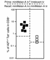
Recall immunizations with miniMelan-A lentivector 60 days after primary immunization with either miniMelan-A lentivector or an irrelevant lentivector. Indicated is the proportion of Melan-A–specific CD8+ cells obtained at the peak of recall response of all individual mice tested (dashed lines indicate the average levels of ELA26–35-specific cells present before recall immunization).
Figure 6
Figure 6
Influence of CD4+ T cells on induction of CD8+ T cell responses by lentivector vaccine. Mice depleted of CD4+ T cells or nondepleted were vaccinated with lentivector vaccine. Indicated are the T cell responses at peak response of groups of mice in the Cw3 model vaccinated with Cw3 cDNA lv and the Melan-A/A2/Kb antigen model vaccinated with ELA26–35 minigene lentivector (x = average, n = number of mice tested per condition). ΔCD4, mice depleted of CD4+ T cells; untr, nondepleted mice.

Similar articles

Cited by

References

    1. Zitvogel L, et al. Therapy of murine tumors with tumor peptide–pulsed dendritic cells: dependence on T cells, B7 costimulation, and T helper cell 1–associated cytokines. J. Exp. Med. 1996;183:87–97. - PMC - PubMed
    1. Nestle FO, et al. Vaccination of melanoma patients with peptide- or tumor lysate–pulsed dendritic cells. Nat. Med. 1998;4:328–332. - PubMed
    1. Thurner B, et al. Vaccination with mage-3A1 peptide-pulsed mature, monocyte-derived dendritic cells expands specific cytotoxic T cells and induces regression of some metastases in advanced stage IV melanoma. J. Exp. Med. 1999;190:1669–1678. - PMC - PubMed
    1. Brossart P, Goldrath AW, Butz EA, Martin S, Bevan MJ. Virus-mediated delivery of antigenic epitopes into dendritic cells as a means to induce CTL. J. Immunol. 1997;158:3270–3276. - PubMed
    1. Song W, et al. Dendritic cells genetically modified with an adenovirus vector encoding the cDNA for a model antigen induce protective and therapeutic antitumor immunity. J. Exp. Med. 1997;186:1247–1256. - PMC - PubMed

Publication types

MeSH terms