Mice deficient in alpha-actinin-4 have severe glomerular disease
- PMID: 12782671
- PMCID: PMC156110
- DOI: 10.1172/JCI17988
Mice deficient in alpha-actinin-4 have severe glomerular disease
Abstract
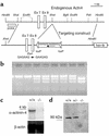
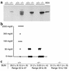
Dominantly inherited mutations in ACTN4, which encodes alpha-actinin-4, cause a form of human focal and segmental glomerulosclerosis (FSGS). By homologous recombination in ES cells, we developed a mouse model deficient in Actn4. Mice homozygous for the targeted allele have no detectable alpha-actinin-4 protein expression. The number of homozygous mice observed was lower than expected under mendelian inheritance. Surviving mice homozygous for the targeted allele show progressive proteinuria, glomerular disease, and typically death by several months of age. Light microscopic analysis shows extensive glomerular disease and proteinaceous casts. Electron microscopic examination shows focal areas of podocyte foot-process effacement in young mice, and diffuse effacement and globally disrupted podocyte morphology in older mice. Despite the widespread distribution of alpha-actinin-4, histologic examination of mice showed abnormalities only in the kidneys. In contrast to the dominantly inherited human form of ACTN4-associated FSGS, here we show that the absence of alpha-actinin-4 causes a recessive form of disease in mice. Cell motility, as measured by lymphocyte chemotaxis assays, was increased in the absence of alpha-actinin-4. We conclude that alpha-actinin-4 is required for normal glomerular function. We further conclude that the nonsarcomeric forms of alpha-actinin (alpha-actinin-1 and alpha-actinin-4) are not functionally redundant. In addition, these genetic studies demonstrate that the nonsarcomeric alpha-actinin-4 is involved in the regulation of cell movement.
Figures


Similar articles
-
Focal and segmental glomerulosclerosis in mice with podocyte-specific expression of mutant alpha-actinin-4.J Am Soc Nephrol. 2003 May;14(5):1200-11. doi: 10.1097/01.asn.0000059864.88610.5e. J Am Soc Nephrol. 2003. PMID: 12707390
-
Alpha-actinin-4-mediated FSGS: an inherited kidney disease caused by an aggregated and rapidly degraded cytoskeletal protein.PLoS Biol. 2004 Jun;2(6):e167. doi: 10.1371/journal.pbio.0020167. Epub 2004 Jun 15. PLoS Biol. 2004. PMID: 15208719 Free PMC article.
-
Mice with podocyte-specific overexpression of wild type alpha-actinin-4 are healthy controls for K256E-alpha-actinin-4 mutant transgenic mice.Transgenic Res. 2010 Apr;19(2):285-9. doi: 10.1007/s11248-009-9305-9. Epub 2009 Jul 8. Transgenic Res. 2010. PMID: 19585264
-
The genetic basis of FSGS and steroid-resistant nephrosis.Semin Nephrol. 2003 Mar;23(2):141-6. doi: 10.1053/snep.2003.50014. Semin Nephrol. 2003. PMID: 12704574 Review.
-
[A genetic viewpoint of focal glomerular sclerosis: fom genes to glomerular pathophysiology [corrected]].G Ital Nefrol. 2003 Jul-Aug;20(4):356-67. G Ital Nefrol. 2003. PMID: 14523896 Review. Italian.
Cited by
-
The carboxyl tail of alpha-actinin-4 regulates its susceptibility to m-calpain and thus functions in cell migration and spreading.Int J Biochem Cell Biol. 2013 Jun;45(6):1051-63. doi: 10.1016/j.biocel.2013.02.015. Epub 2013 Mar 1. Int J Biochem Cell Biol. 2013. PMID: 23466492 Free PMC article.
-
TRPC6 in glomerular health and disease: what we know and what we believe.Semin Cell Dev Biol. 2006 Dec;17(6):667-74. doi: 10.1016/j.semcdb.2006.11.003. Epub 2006 Nov 20. Semin Cell Dev Biol. 2006. PMID: 17116414 Free PMC article. Review.
-
HAMLET binding to α-actinin facilitates tumor cell detachment.PLoS One. 2011 Mar 8;6(3):e17179. doi: 10.1371/journal.pone.0017179. PLoS One. 2011. PMID: 21408150 Free PMC article.
-
Actinin-4 in keratinocytes regulates motility via an effect on lamellipodia stability and matrix adhesions.FASEB J. 2013 Feb;27(2):546-56. doi: 10.1096/fj.12-217406. Epub 2012 Oct 19. FASEB J. 2013. PMID: 23085994 Free PMC article.
-
The next generation of therapeutics for chronic kidney disease.Nat Rev Drug Discov. 2016 Aug;15(8):568-88. doi: 10.1038/nrd.2016.67. Epub 2016 May 27. Nat Rev Drug Discov. 2016. PMID: 27230798 Free PMC article. Review.
References
-
- Ichikawa I, Fogo A. Focal segmental glomerulsoclerosis. Pediatr. Nephrol. 1996;10:374–391. - PubMed
-
- Kaplan JM, et al. Mutations in ACTN4, encoding alpha-actinin-4, cause familial focal segmental glomerulosclerosis. Nat. Genet. 2000;24:251–256. - PubMed
-
- Kestila M, et al. Positionally cloned gene for a novel glomerular protein — nephrin — is mutated in congenital nephrotic syndrome. Mol. Cell. 1998;1:575–582. - PubMed
-
- Boute N, et al. NPHS2, encoding the glomerular protein podocin, is mutated in autosomal recessive steroid-resistant nephrotic syndrome. Nat. Genet. 2000;24:349–354. - PubMed
-
- Shih NY, et al. Congenital nephrotic syndrome in mice lacking CD2-associated protein. Science. 1999;286:312–315. - PubMed
Publication types
MeSH terms
Substances
Grants and funding
LinkOut - more resources
Full Text Sources
Other Literature Sources
Medical
Molecular Biology Databases
Miscellaneous

