. 2003 Jun;111(11):1703-13.
doi: 10.1172/JCI16921.

The voltage-gated Kv1.3 K(+) channel in effector memory T cells as new target for MS

Affiliations

The voltage-gated Kv1.3 K(+) channel in effector memory T cells as new target for MS

Heike Wulff et al. J Clin Invest. 2003 Jun.

Erratum in

  • J Clin Invest. 2003 Jul;112(2):298

Abstract

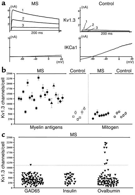
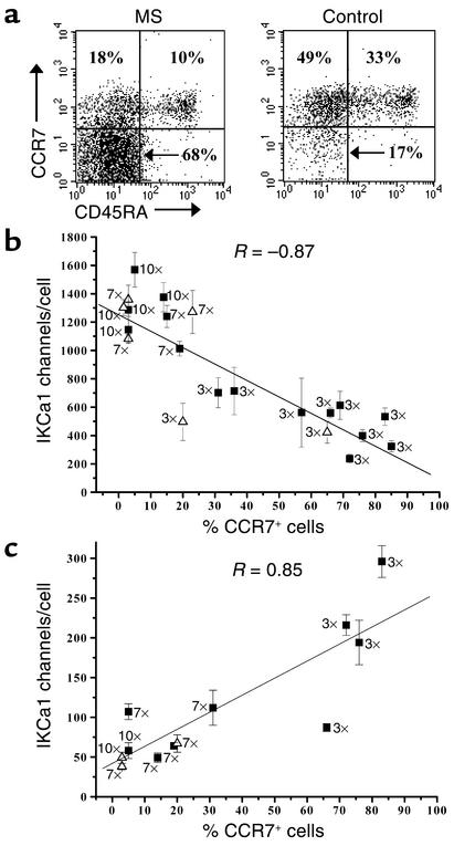
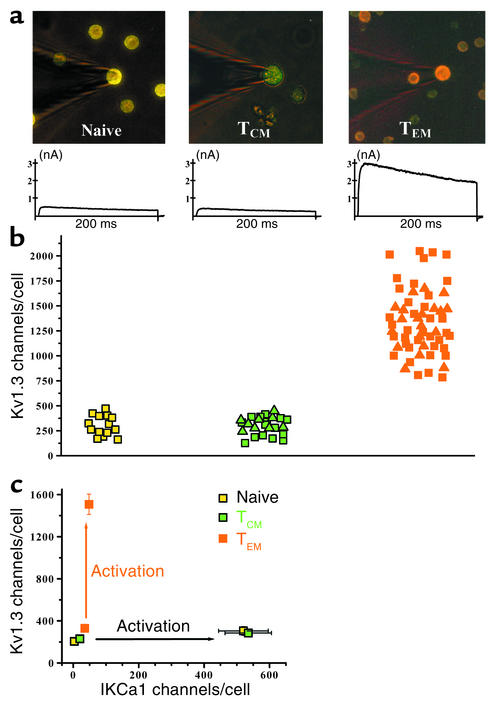
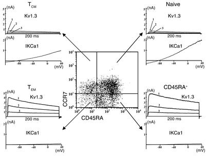
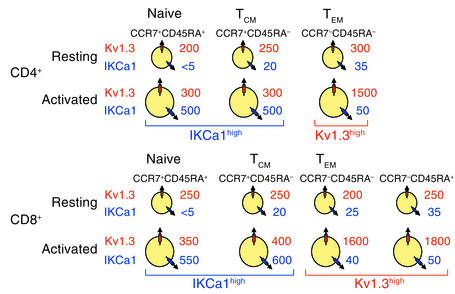
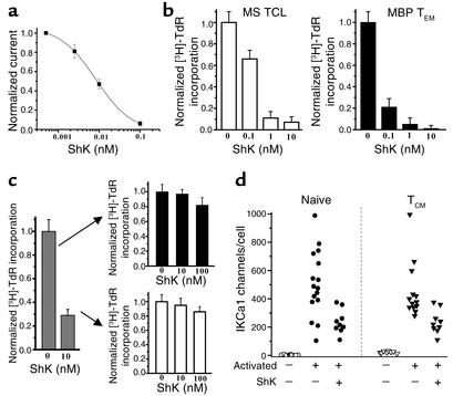
Through a combination of fluorescence microscopy and patch-clamp analysis we have identified a striking alteration in K(+) channel expression in terminally differentiated human CCR7(-)CD45RA(-) effector memory T lymphocytes (T(EM)). Following activation, T(EM) cells expressed significantly higher levels of the voltage-gated K(+) channel Kv1.3 and lower levels of the calcium-activated K(+) channel IKCa1 than naive and central memory T cells (T(CM)). Upon repeated in vitro antigenic stimulation, naive cells differentiated into Kv1.3(high)IKCa1(low) T(EM) cells, and the potent Kv1.3-blocking sea anemone Stichodactyla helianthus peptide (ShK) suppressed proliferation of T(EM) cells without affecting naive or T(CM) lymphocytes. Thus, the Kv1.3(high)IKCa1(low) phenotype is a functional marker of activated T(EM) lymphocytes. Activated myelin-reactive T cells from patients with MS exhibited the Kv1.3(high)IKCa1(low) T(EM) phenotype, suggesting that they have undergone repeated stimulation during the course of disease; these cells may contribute to disease pathogenesis due to their ability to home to inflamed tissues and exhibit immediate effector function. The Kv1.3(high)IKCa1(low) phenotype was not seen in glutamic acid decarboxylase, insulin-peptide or ovalbumin-specific and mitogen-activated T cells from MS patients, or in myelin-specific T cells from healthy controls. Selective targeting of Kv1.3 in T(EM) cells may therefore hold therapeutic promise for MS and other T cell-mediated autoimmune diseases.


Figures

Figure 1
Figure 1
Myelin antigen–activated T cells from MS patients express the distinctive Kv1.3highIKCa1lowphenotype. (a) Kv1.3 and IKCa1 currents in TCLs from MS patients and controls measured 48 hours after activation with MBP. The numbers 1, 2, and 3 in the Kv1.3 traces represent traces after the first, second, and third depolarizing 200-ms pulse; the pulse interval is 1 second. (b) Kv1.3 channel number/cell in myelin antigen–activated TCLs (left) or mitogen-activated T cells (right) from MS patients or controls. Each data point constitutes the mean ± SEM of 20–30 cells from two to seven independent TCLs. Myelin antigens used were MBP (filled and open squares), MOG (filled and open triangles), and PLP (filled diamonds). Filled symbols, MSpatients; open symbols, control subjects. Mitogens used were soluble anti-CD3 mAb (50 ng/ml) and PMA (10 nM) plus ionomycin (175 nM) (data for both mitogens are grouped together). Filled circles, MS patients; open circles, controls. (c) Kv1.3 channel number/cell in MS patient T cells activated for 48 hours with the control antigens GAD65, insulin peptide, and ovalbumin. Due to the paucity of cells in these TCLs, IKCa1 expression was measured in only two to three cells per TCL.
Figure 2
Figure 2
Cells with altered channel expression are phenotypically effector memory T cells. (a) Flow cytometry profiles showing CD45RA and CCR7 expression in MBP-specific TCLs from an MS patient and a control subject. (b) Kv1.3 channel number/cell versus percentage of CCR7+ cells in control TCLs stimulated three, seven, and ten times with MBP (filled squares) or TT (open triangles). The number of antigenic stimulations is shown in the figure next to each symbol. Twenty or more cells were studied from each sample. (c) IKCa1 channel number/cell versus percentage of CCR7+cells in the same TCLs as in b. Fewer data points are included in the IKCa1 plot because the number of cells in some myelin antigen-specific TCL samples were not adequate to measure both Kv1.3 and IKCa1 expression in 20 cells each.
Figure 3
Figure 3
Kv1.3highIKCa1low phenotype: an exclusive functional marker for activated effector memory T cells. (a) Fluorescent immunostained images of patch-clamped CD4 T cell subsets from control subjects showing naive CCR7+CD45RA+ (left), TCM CCR7+CD45RAcells (middle), and TEM CCR7+CD45RA+cells (right, counterstained with antihuman CD4 mAb). Representative Kv1.3 currents from each cell type are shown below the respective images. (b) Kv1.3 channel number/cell in naive (yellow squares), TCM (MBP-stimulated, green squares; TT-stimulated, green triangles) and TEM (MBP-stimulated, orange squares; TT-stimulated, orange triangles) T cells. Activated naive CD4+ cells were identified in the peripheral blood of controls 48 hours following stimulation with anti-CD3 mAb. Activated TCM and TEM cells were identified in the same preparations of control TCLs (stimulated ten times with either MBP or TT) 48 hours after stimulation with the appropriate antigen. (c) Kv1.3 versus IKCa1 channel number per cell in the three populations before and after activation. Each data point is the mean ± SEM channel number in 20–50 cells.
Figure 4
Figure 4
The Kv1.3highIKCa1low phenotype is also found in activated CD8+ effector memory T cells. Flow cytometry profile showing CD45RA and CCR7 expression in activated CD8+ T cells. Representative Kv1.3 and IKCa1 currents from the naive (top right), TCM (top left), TEM (bottom left), and CD45RA+ (bottom right, CCR7CD45RA+) subsets are shown next to the FACS quadrants.
Figure 5
Figure 5
Diagram of average Kv1.3 and IKCa1 channel numbers per cell in the three CD4+ and four CD8+ CCR7/CD45RA-distinguished T cell subsets in the quiescent and the activated state. Mean channel numbers (n = 10–50 cells for each subset) are shown.
Figure 6
Figure 6
Kv1.3 blockers selectively and persistently suppress MBP-specific TEM cells. (a) ShK potently blocked Kv1.3 in Kv1.3highIKCa1low MBP-specific TEM cells Kd, 9 ± 1 pM). Each data point represents the mean ± SD of three cells. (b) ShK suppressed anti-CD3 mAb–stimulated [3H]TdR incorporation by a Kv1.3highIKCa1low MBP-specific MS patient TCL stimulated three times with MBP (left) and a control TCL stimulated 12 times with MBP (right). (c) ShK (10 nM) partially suppressed anti-CD3 mAb–stimulated [3H]TdR incorporation by normal peripheral blood T lymphocytes (left) but was unable to suppress proliferation when these cells were rechallenged with anti-CD3 mAb. One representative experiment from a total of three experiments is shown. Each experiment was done in triplicate. (d) IKCa1 expression is enhanced in activated naive and TCM cells following anti-CD3 mAb stimulation, despite the presence of a suppressive dose of ShK. Open circles, naive resting; filled circles, naive T cells activated by anti-CD3 mAb in the absence or presence of ShK; open triangles, TCM resting; filled triangles, TCM cells activated by anti-CD3 mAb in the absence or presence of ShK.

References

    1. Allegretta M, Nicklas JA, Sriram S, Albertini RJ. T cells responsive to myelin basic protein in patients with multiple sclerosis. Science. 1990;247:718–721. - PubMed
    1. Lovett-Racke AE, et al. Decreased dependence of myelin basic protein-reactive T cells on CD28-mediated costimulation in multiple sclerosis patients: a marker of activated/memory T cells. J. Clin. Invest. 1998;101:725–730. - PMC - PubMed
    1. Scholz C, Anderson DE, Freeman GJ, Hafler DA. Expansion of autoreactive T cells in multiple sclerosis is independent of exogenous B7 costimulation. J. Immunol. 1998;160:1532–1538. - PubMed
    1. Markovic-Plese S, Cortese I, Wandinger KP, McFarland HF, Martin R. CD4+CD28– costimulation-independent T cells in multiple sclerosis. J. Clin. Invest. 2001;108:1185–1194. doi:10.1172/JCI200112516. - PMC - PubMed
    1. Viglietta V, Kent SC, Orban T, Hafler DA. GAD65-reactive T cells are activated in patients with autoimmune type 1a diabetes. J. Clin. Invest. 2002;109:895–903. doi:10.1172/JCI200214114. - PMC - PubMed

Publication types

MeSH terms