. 2003 Jun;111(11):1757-69.
doi: 10.1172/JCI17993.

Polymorphisms in Chlamydia trachomatis tryptophan synthase genes differentiate between genital and ocular isolates

Affiliations

Polymorphisms in Chlamydia trachomatis tryptophan synthase genes differentiate between genital and ocular isolates

Harlan D Caldwell et al. J Clin Invest. 2003 Jun.

Abstract

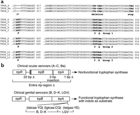
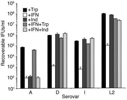
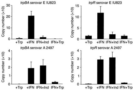
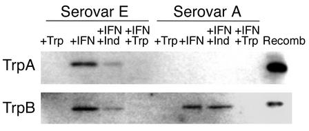
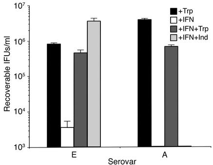
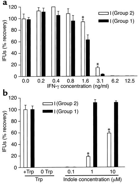
We previously reported that laboratory reference strains of Chlamydia trachomatis differing in infection organotropism correlated with inactivating mutations in the pathogen's tryptophan synthase (trpBA) genes. Here, we have applied functional genomics to extend this work and find that the paradigm established for reference serovars also applies to clinical isolates - specifically, all ocular trachoma isolates tested have inactivating mutations in the synthase, whereas all genital isolates encode a functional enzyme. Moreover, functional enzyme activity was directly correlated to IFN-gamma resistance through an indole rescue mechanism. Hence, a strong selective pressure exists for genital strains to maintain a functional synthase capable of using indole for tryptophan biosynthesis. The fact that ocular serovars (serovar B) isolated from the genital tract were found to possess a functional synthase provided further persuasive evidence of this association. These results argue that there is an important host-parasite relationship between chlamydial genital strains and the human host that determines organotropism of infection and the pathophysiology of disease. We speculate that this relationship involves the production of indole by components of the vaginal microbial flora, allowing chlamydiae to escape IFN-gamma-mediated eradication and thus establish persistent infection.


Figures

Figure 1
Figure 1
(a) Alignment of partial sequences from the trpA gene from the14 human C. trachomatis reference serovars. A ClustalW alignment of the nucleotide regions containing sequence polymorphisms is illustrated. As compared with genital serovars, ocular serovars have a three-base (nucleotides 408–410) deletion that results in the loss of a phenylalanine. The various serovars have been grouped, in accordance with their nucleotide mutational “hot-spot” sequence. The ocular serovars have a single-base deletion (nucleotide 528) resulting in a nonfunctional truncated TrpA protein. Genital serovar specific missense mutations (nucleotides 530 and 532) that result in amino acid changes in loop 6 of TrpA are shown below the nucleotide sequence. See Fehlner-Gardiner et al. (16) for details. (b) Schematic summary of the tryptophan synthase inactivating mutations identified in clinical ocular serovars and the missense mutations identified in the clinical genital serovars.
Figure 2
Figure 2
Effect of indole or tryptophan on the growth of C. trachomatis reference serovars A, D, I, and L2 cultured in the presence of IFN-γ. HeLa cell monolayers were infected with EBs at an moi of 3–5 IFUs/cell. For serovar L2 but not A, D, or I, HeLa cell monolayers were pretreated with IFN-γ (5 ng/ml) for 24 hours before infection. Infected HeLa cells were cultured in the presence of complete DMEM-10 (+Trp), complete DMEM-10 plus 5 ng/ml IFN-γ (+IFN), complete DMEM-10 plus IFN-γ and 100 μM indole (IFN+Ind), and complete DMEM-10 plus IFN-γ and supplemented with 1 g/l tryptophan (IFN+Trp). After 48 hours for serovar L2 and 72 hours for serovars A, D and I, infected cells and culture supernatants were collected and used to infect a new HeLa cell monolayer for enumeration of recoverable IFUs. Data are presented as IFUs (log10) and represent the means ± SD of triplicate determinations.
Figure 3
Figure 3
RT-PCR quantitation of trpBA and trpR mRNA isolated from HeLa cell monolayers infected with C. trachomatis ocular (serovar A 2497) and genital (serovars E IU823) clinical isolates cultured in the presence of IFN-γ. Culture and chlamydiae infection conditions were as described in the legend to Figure 2. Infected HeLa cells were cultured in the presence of complete DMEM-10 (+Trp), complete DMEM-10 plus 5 ng/ml IFN-γ (+IFN), complete DMEM-10 plus IFN-γ and 100 μM indole (IFN+Ind), and complete DMEM-10 plus IFN-γ and supplemented with 1 g/l tryptophan (IFN+Trp). RNA was isolated 36 hours after infection. The RNA was reverse transcribed using random hexamer primers, and the cDNA was used for quantitative PCR amplifications with primers specific for trpBA or trpR. Quantitative PCR reactions were carried out using the LightCycler and SYBR Green I as the fluorophore. See Methods for details.
Figure 4
Figure 4
Western blot analysis of TrpA and TrpB expression in HeLa cell monolayers infected with C. trachomatis ocular clinical isolate serovar A 2497 or genital clinical isolate serovar E IU823 cultured in the presence of IFN-γ. Infected HeLa cells were cultured in the presence of complete DMEM-10 (+Trp), complete DMEM-10 plus 5 ng/ml IFN-γ (+IFN), complete DMEM-10 plus IFN-γ and 100 μM indole (IFN+Ind), and complete DMEM-10 plus IFN-γ and supplemented with 1 g/l tryptophan (IFN+Trp). Infected cells were harvested 48 hours after infection and sonicated, and chlamydial particles were semipurified over a 30% MD-76 cushion. The resulting pellet was lysed in Laemmli sample buffer, and proteins were separated by SDS-PAGE (12%) and then transferred to nitrocellulose. Proteins were detected using polyclonal antibodies raised against recombinant L2 TrpB or TrpA. The respective recombinant proteins (Recomb) were included as positive controls.
Figure 5
Figure 5
Effect of indole or tryptophan on the growth of C. trachomatis ocular (serovar A 2497) and genital (serovar E IU823) clinical isolates cultured in the presence of IFN-γ. Culture and chlamydiae infection conditions were as described in the legend to Figure 2. Infected HeLa cells were cultured in the presence of complete DMEM-10 (+Trp), complete DMEM-10 plus 5 ng/ml IFN-γ (+IFN), complete DMEM-10 plus IFN-γ and 100 μM indole (IFN+Ind), and complete DMEM-10 plus IFN-γ and supplemented with 1 g/l tryptophan (IFN+Trp). After 72 hours, cultures were harvested and recoverable IFUs were enumerated. Data are presented as IFUs (log10) and represent the means ± SD of triplicate determinations.
Figure 6
Figure 6
IFN-γ sensitivity and indole rescue of paired I serovariants as a function of the Group 1/2 polymorphism in trpA. (a) Chlamydial isolates were grown in the presence of a range of IFN-γ concentrations in complete DMEM-10 in the absence of cyclohexamide on HeLa monolayers at an moi of 0.1. After 48 hours, cultures were harvested and recoverable IFUs were enumerated. Graphed values represent the percent recovery from each concentration of IFN-γ based on the 0 IFN-γ well of each isolate equaling 100% recovery. (b) Chlamydial isolates were grown in complete DMEM-10 or DMEM-10(-Trp) on HeLa monolayers at an moi of 0.5. Tryptophan minus wells were rescued 24 hours after infection with three different indole concentrations. After a further 48 hours, cultures were harvested and recoverable IFUs were enumerated. Graphed values represent the percent recovery from each indole concentration based on the plus tryptophan well of each serovar equaling 100% recovery. The differences in recovery were significant (P < 0.05) at the 1 and 10 μM indole concentrations.
Figure 7
Figure 7
Indirect fluorescent antibody staining of C. trachomatis–infected HeLa cells with mAbs specific to ompA. Note that the B genital isolate reacts with the same specificity as the ocular B laboratory reference strain. Strains tested are shown across the top of the figure; mAbs used to stain infected cells are shown on the left hand side of the figure. HeLa cells were infected with an moi of 0.2. Monolayers were fixed in methanol at 36–48 hours after infection. Chlamydiae were detected by reacting fixed cells with individual mAbs followed by staining with an FITC goat anti-mouse Ig antiserum. The specificity of the mAbs are as follows: BB6 (serovar B), JG9 (serovar D), BB5 (serovars B and D), and L2I-10 (all C. trachomatis serovars, A-L3).
Figure 8
Figure 8
(a) Western blot analysis of TrpA and TrpB expression in HeLa cell monolayers infected with a C. trachomatis clinical B isolate. Cell culture conditions, harvesting, chlamydial protein sample preparation, and Western blot procedure were as described in the legend to Figure 4. (b) Effect of indole or tryptophan on the growth of C. trachomatis genital serovar B clinical isolate cultured in the presence of IFN-γ. Cell culture conditions, harvesting at 72 hours after infection, and enumeration of recoverable infectious chlamydial EBs were as described in the legend to Figure 2. Data are presented as IFUs (log10) and represent the means ± SD of triplicate determinations.

Comment in

References

    1. Moulder JW. Interaction of chlamydiae and host cells in vitro. Microbiol. Rev. 1991;55:143–190. - PMC - PubMed
    1. Schachter, J. 1999. Infection and disease epidemiology. American Society for Microbiology Press. Washington, DC, USA. 139–170.
    1. Stephens, R.S. 1999. Genomic autobiographies of Chlamydiae. American Society for Microbiology Press. Washington, DC, USA. 9–27.
    1. An BB, Adamis AP. Chlamydial ocular diseases. Int. Ophthalmol. Clin. 1998;38:221–230. - PubMed
    1. Mabey D, Bailey R. Eradication of trachoma worldwide. Br. J. Ophthalmol. 1999;83:1261–1263. - PMC - PubMed

Publication types

MeSH terms