. 2003 Jun;111(11):1783-91.
doi: 10.1172/JCI16100.

The alpha(1A/C)- and alpha(1B)-adrenergic receptors are required for physiological cardiac hypertrophy in the double-knockout mouse

Affiliations

The alpha(1A/C)- and alpha(1B)-adrenergic receptors are required for physiological cardiac hypertrophy in the double-knockout mouse

Timothy D O'Connell et al. J Clin Invest. 2003 Jun.

Abstract

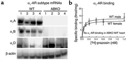
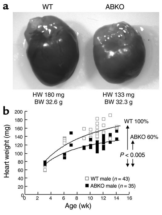
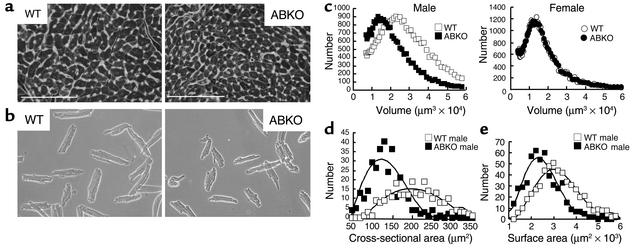
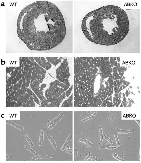
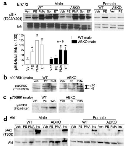
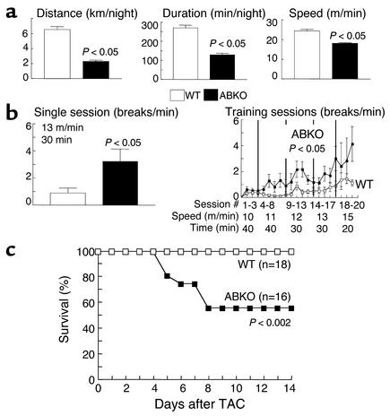
Catecholamines and alpha(1)-adrenergic receptors (alpha(1)-ARs) cause cardiac hypertrophy in cultured myocytes and transgenic mice, but heart size is normal in single KOs of the main alpha(1)-AR subtypes, alpha(1A/C) and alpha(1B). Here we tested whether alpha(1)-ARs are required for developmental cardiac hypertrophy by generating alpha(1A/C) and alpha(1B) double KO (ABKO) mice, which had no cardiac alpha(1)-AR binding. In male ABKO mice, heart growth after weaning was 40% less than in WT, and the smaller heart was due to smaller myocytes. Body and other organ weights were unchanged, indicating a specific effect on the heart. Blood pressure in ABKO mice was the same as in WT, showing that the smaller heart was not due to decreased load. Contractile function was normal by echocardiography in awake mice, but the smaller heart and a slower heart rate reduced cardiac output. alpha(1)-AR stimulation did not activate extracellular signal-regulated kinase (Erk) and downstream kinases in ABKO myocytes, and basal Erk activity was lower in the intact ABKO heart. In female ABKO mice, heart size was normal, even after ovariectomy. Male ABKO mice had reduced exercise capacity and increased mortality with pressure overload. Thus, alpha(1)-ARs in male mice are required for the physiological hypertrophy of normal postnatal cardiac development and for an adaptive response to cardiac stress.


Figures

Figure 1
Figure 1
α1-AR subtype mRNA levels and receptor binding in hearts from ABKO and WT mice. (a) α1-AR subtype mRNAs by RPA. RNA was from ventricles of 10-week-old male mice. (b) α1-AR saturation binding. [3H]-prazosin was used with ventricular membranes from 10-week-old male and female mice; n = 4 of each sex and genotype.
Figure 2
Figure 2
Heart growth in male ABKO and WT mice. (a) Typical hearts at 12 weeks. Twelve-week-old male mice had the wet HWs and BWs shown. (b) HW versus age. Wet HW in male mice from weaning (3 weeks) to young adulthood (14 weeks) is plotted versus age. Curves were fit using nonlinear regression (F = 17.81, P < 0.005). Heart growth after weaning in the ABKO mice was only 60% of that in WT; BW was the same as in WT (not shown).
Figure 3
Figure 3
Myocyte size in ABKO and WT mice. (a) Ventricular cross sections. LV coronal sections from 10- to 11-week-old male mice were stained with fluorescein-conjugated wheat germ agglutinin for sarcolemmal membranes and with Hoechst 33342 for nuclei. Nuclei between myocytes are in nonmyocytes. ×40. Scale bar, 100 μm. (b) Isolated myocytes. Myocytes were from hearts of 9- to 10-week-old male mice. ×20, phase contrast. (c) Myocyte volume. Myocyte volume by Coulter Multisizer is plotted versus myocyte number for representative hearts of each genotype and sex. (d) Myocyte cross-sectional area. Cross-sectional area in myocytes with a central nucleus, as in a, is plotted versus myocyte number (110–190 cells in each of two male hearts for each genotype). (e) Myocyte surface area. Surface areas of isolated myocytes as in b are plotted versus myocyte number (160–190 cells in each of three male hearts for each genotype). Group data are in Table 1.
Figure 4
Figure 4
Heart and myocyte size in congenic 10- to 12-week-old male ABKO and WT mice. (a) Whole-heart cross sections. Frozen sections of whole heart were stained with hematoxylin. ×2.5. The area of the ABKO section is 70% of that of the WT. (b) Ventricular cross sections. Hearts were fixed in diastole, and coronal sections of the left ventricle were stained with 1% toluidine blue. Small clear areas are capillaries that mark myocyte borders. ×40. (c) Isolated myocytes. ×20, phase contrast. Myocyte surface area was 3,259 ± 1,024 μm2 in WT mice (n = 45) and 2,454 ± 812 μm2 in ABKO mice (n = 41, 75% of WT, P < 0.05).
Figure 5
Figure 5
Hypertrophic signaling in myocytes. Cultured myocytes from hearts of 10- to 11-week-old congenic WTand ABKO mice were treated for 15 minutes with PE (20 μM, plus 2 μM timolol); phorbol 12-myristate, 13-acetate (PMA, 100 nM); sorbitol (Sor, 1 M); endothelin (ET, 10 nM); insulin (Ins, 6 μM); or vehicle. Western blots were done for (a) phospho-Erk1/2 (pErk) and total Erk1/2, (b) phospho-p90RSK (pp90RSK), (c) phospho-p70S6K (pp70S6K), and (d) phospho-Akt (pAkt) and total Akt. In a, the bar graphs summarize Erk1/2 data from six hearts in each group. In b, a nonspecific (NS) band is indicated with anti–pp90RSK. In d, a second pAkt antibody (S473) gave identical results (not shown). Total p90RSK (b) and total p70S6K (c) were the same in WT and ABKO mice (not shown). *P < 0.05 vs. Veh.
Figure 6
Figure 6
Response of the ABKO mice to stress. Ten- to twelve-week-old male ABKO and WT mice were used. (a) Exercise: free wheel running. Mice were given access to a running wheel for 12 hours on each of 30 consecutive nights, and distance, duration, and speed were recorded by a chronometer attached to the wheel (n = 4–5 mice each group). (b) Exercise: motorized treadmill. Mice were placed on a motorized treadmill set at the speed and for the duration indicated, and the number of times the motivational bar was touched, indicating failure to maintain treadmill speed, was recorded as breaks per minute. The left panel shows a single session (n = 8–9 mice per group), and the right panel shows 20 consecutive daily training sessions (n = 9–10). (c) TAC was done in congenic mice, and survival was recorded over 14 days.

References

    1. Rakusan, K. 1984. Cardiac growth, maturation and aging. In Growth of the heart in health and disease. R. Zak, editor. Raven Press. New York, New York, USA. 131–164.
    1. Simpson PC, Kariya K, Karns LR, Long CS. The α1-adrenergic receptor in left ventricular hypertrophy. Journal of Vascular Medicine and Biology. 1990;2:236–246.
    1. Rapacciuolo A, et al. Important role of endogenous norepinephrine and epinephrine in the development of in vivo pressure-overload cardiac hypertrophy. J. Am. Coll. Cardiol. 2001;38:876–882. - PubMed
    1. Thomas SA, Matsumoto AM, Palmiter RD. Noradrenaline is essential for mouse fetal development. Nature. 1995;374:643–646. - PubMed
    1. Simpson P. Norepinephrine-stimulated hypertrophy of cultured rat myocardial cells is an alpha1-adrenergic response. J. Clin. Invest. 1983;72:732–738. - PMC - PubMed

Publication types

MeSH terms

Substances