

An overview of the eye in diabetes
- PMID: 12782689
- PMCID: PMC539505
- DOI: 10.1177/014107680309600603
An overview of the eye in diabetes
Abstract
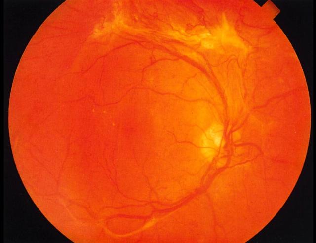
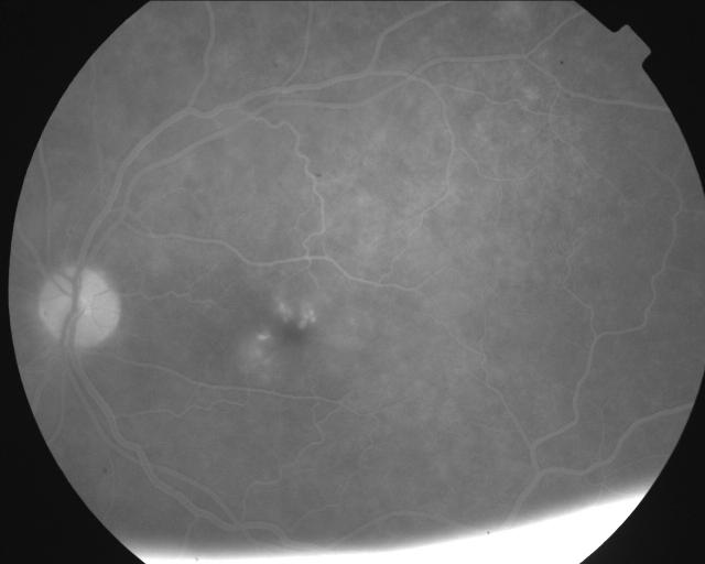
The three papers in this symposium are based on presentations to an RSM meeting on the Diabetic Eye, held on 9 April 2003. The matter is particularly topical because the National Service Framework for Diabetes calls for a high-quality retinal screening programme. After a review of the various ophthalmic conditions likely to be encountered in diabetic patients (A Negi, S A Vernon) we proceed to the most important, diabetic retinopathy, with a discussion of screening methods (D M Squirrell, J F Talbot) and an account of laser treatments (J G F Dowler). Colour versions of the clinical photographs are available online [www.jrsm.org]. Publication was coordinated by Professor Susan Lightman, of Moorfields Eye Hospital, London, UK.
Figures
Similar articles
-
Ocular manifestations of diabetes mellitus.Postgrad Med. 1980 Oct;68(4):143-5, 148-9, 153-7. doi: 10.1080/00325481.1980.11715568. Postgrad Med. 1980. PMID: 7422598 No abstract available.
-
Diabetes and the eye.Clin Symp. 1984;36(4):2-32. Clin Symp. 1984. PMID: 6085831 No abstract available.
-
Ocular complications of diabetes.Clin Symp. 1992;44(1):2-32. Clin Symp. 1992. PMID: 1559315 Review. No abstract available.
-
Diabetic eye disease: a primary care perspective.South Med J. 1996 May;89(5):463-70. doi: 10.1097/00007611-199605000-00002. South Med J. 1996. PMID: 8638169 Review.
-
Noninfectious ocular complications of AIDS.Int Ophthalmol Clin. 1989 Summer;29(2):95-7. doi: 10.1097/00004397-198902920-00006. Int Ophthalmol Clin. 1989. PMID: 2654050 Review. No abstract available.
Cited by
-
Optical Coherence Tomography Angiography in Diabetes and Diabetic Retinopathy.J Clin Med. 2020 Jun 3;9(6):1723. doi: 10.3390/jcm9061723. J Clin Med. 2020. PMID: 32503234 Free PMC article. Review.
-
Vascular adhesion protein-1 regulates leukocyte transmigration rate in the retina during diabetes.Exp Eye Res. 2009 Nov;89(5):774-81. doi: 10.1016/j.exer.2009.07.010. Epub 2009 Jul 25. Exp Eye Res. 2009. PMID: 19635478 Free PMC article.
-
Atorvastatin-Eluting Contact Lenses: Effects of Molecular Imprinting and Sterilization on Drug Loading and Release.Pharmaceutics. 2021 Apr 22;13(5):606. doi: 10.3390/pharmaceutics13050606. Pharmaceutics. 2021. PMID: 33922123 Free PMC article.
-
Enhanced wound healing, kinase and stem cell marker expression in diabetic organ-cultured human corneas upon MMP-10 and cathepsin F gene silencing.Invest Ophthalmol Vis Sci. 2013 Dec 17;54(13):8172-80. doi: 10.1167/iovs.13-13233. Invest Ophthalmol Vis Sci. 2013. PMID: 24255036 Free PMC article.
-
Exploring exercise-driven inhibition of pyroptosis: novel insights into treating diabetes mellitus and its complications.Front Endocrinol (Lausanne). 2023 Oct 4;14:1230646. doi: 10.3389/fendo.2023.1230646. eCollection 2023. Front Endocrinol (Lausanne). 2023. PMID: 37859981 Free PMC article. Review.
References
-
- Chief Medical Officer. Annual Report of the Chief Medical Officer of the Department of Health for the Year 1997. London: Stationery Office, 1998
-
- Evans J. Causes of Blindness and Partial Sight in England and Wales 1990-91. London: HMSO
-
- McLeod BK, Thompson JR, Rosenthal AR. The prevalence of retinopathy in the insulin-requiring diabetic patients of an English country town. Eye 1988;2: 424-30 - PubMed
-
- Klein R, Klein BE, Moss SE, et al. The Wisconsin Epidemiologic Study of Diabetic Retinopathy. III. Prevalence and risk of diabetic retinopathy when age at diagnosis is 30 or more years. Arch Ophthalmol 1984;102: 527-32 - PubMed
-
- Hawthorne K, Mello M, Tomlinson S. Cultural and religious influences in diabetes care in Great Britain. Diabet Med 1993;10: 8-12 - PubMed
Publication types
MeSH terms
LinkOut - more resources
Full Text Sources
Medical
Miscellaneous
