. 2003 Jun 2;197(11):1405-16.
doi: 10.1084/jem.20030109.

Vaccine immunity to pathogenic fungi overcomes the requirement for CD4 help in exogenous antigen presentation to CD8+ T cells: implications for vaccine development in immune-deficient hosts

Affiliations

Vaccine immunity to pathogenic fungi overcomes the requirement for CD4 help in exogenous antigen presentation to CD8+ T cells: implications for vaccine development in immune-deficient hosts

Marcel Wuthrich et al. J Exp Med. .

Abstract

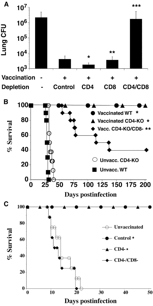
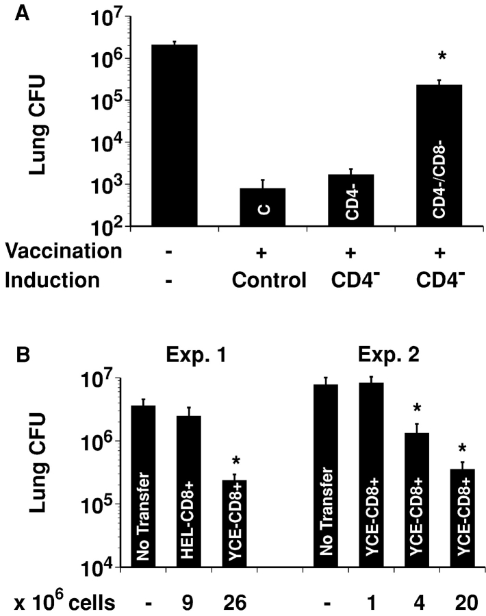
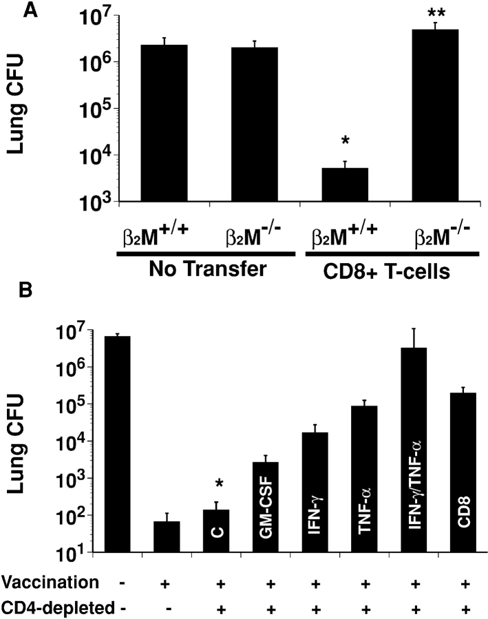
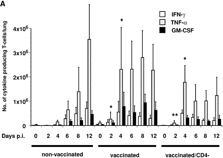
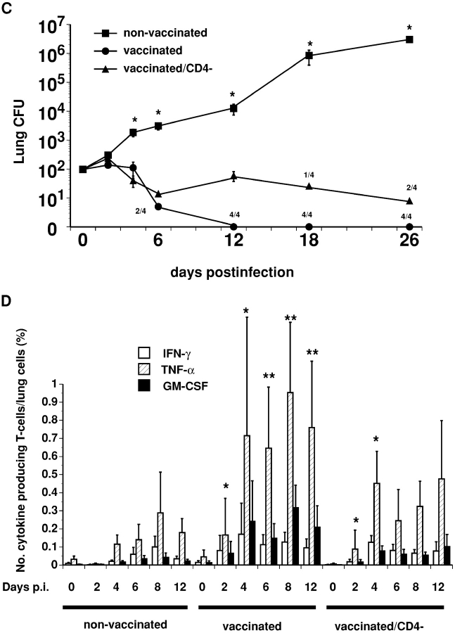
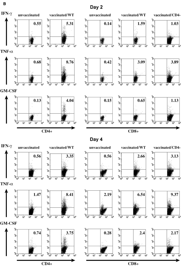
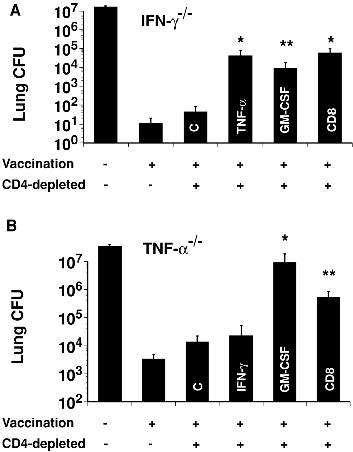
Systemic fungal infections with primary and opportunistic pathogens have become increasingly common and represent a growing health menace in patients with AIDS and other immune deficiencies. T lymphocyte immunity, in particular the CD4+ Th 1 cells, is considered the main defense against these pathogens, and their absence is associated with increased susceptibility. It would seem illogical then to propose vaccinating these vulnerable patients against fungal infections. We report here that CD4+ T cells are dispensable for vaccine-induced resistance against experimental fungal pulmonary infections with two agents, Blastomyces dermatitidis an extracellular pathogen, and Histoplasma capsulatum a facultative intracellular pathogen. In the absence of T helper cells, exogenous fungal antigens activated memory CD8+ cells in a major histocompatibility complex class I-restricted manner and CD8+ T cell-derived cytokines tumor necrosis factor alpha, interferon gamma, and granulocyte/macrophage colony-stimulating factor-mediated durable vaccine immunity. CD8+ T cells could also rely on alternate mechanisms for robust vaccine immunity, in the absence of some of these factors. Our results demonstrate an unexpected plasticity of immunity in compromised hosts at both the cellular and molecular level and point to the feasibility of developing vaccines against invasive fungal infections in patients with severe immune deficiencies, including those with few or no CD4+ T cells.


Figures

Figure 1.
Figure 1.
Role of CD4+ and CD8+ T cells during induction of vaccine immunity. (A) Lung CFU analysis. Vaccinated mice (10–12/group) were depleted of T cell subsets during vaccination and infection. Mice were infected with 2 × 10³ yeast and analyzed for lung CFU 14–16 d after infection. Panel A shows an average of two independent experiments. *, P = 0.1983 vs. vaccinated control (rat IgG treated), **, P = 0.9825, vs. control, ***, P = 0.7 vs. unvaccinated mice. (B) Survival of vaccinated, CD4+ T cell knockout mice and wild-type mice (10–11/group). Vaccinated CD4-KO/CD8- mice were depleted of CD8+ T cells after vaccination and throughout infection. *, P < 0.0001 vs. unvaccinated mice; **, P = 0.0077 vs. vaccinated CD4-KO mice. (C) CD8+ T cells mediate vaccine immunity to H. capsulatum. Anti-CD4-/anti-CD8 denotes vaccinated CD4-depleted mice that were depleted of CD8+ T cells after vaccination and throughout infection. Eight mice/group were studied. *, P < 0.0002 vs. unvaccinated mice or anti-CD4-/anti-CD8 mAb treated mice.
Figure 2.
Figure 2.
CD8+ T cells mediate resistance in the absence of CD4+ T cells. (A) Depletion of CD8+ T cells impairs resistance. C57BL/6 mice were depleted of CD4+ T cells during vaccination (induction). As indicated in histogram bars, after infection, mice also were depleted of CD4+ cells alone or together with CD8+ T cells. Controls were vaccinated mice treated with rat-IgG (C) or unvaccinated mice. Mice were infected with 2 × 10³ yeast and analyzed for lung CFU 14 d later. *, P < 0.0001 vs. CD4-depleted mice. Data represent an average of two independent experiments (n = 10 mice/group). (B) CD8+ T cells transfer resistance. In experiment 1, naive mice (8/group) received either Blastomyces immune (YCE) CD8+ T cells, no cells, or control HEL-CD8+ T cells. Mice were infected 1d later with 103 yeast, and lung CFU analyzed 2 wk after infection, *, P < 0.001 vs. mice receiving either HEL-CD8+ T cells or no cells. Data show one representative experiment of three independent experiments. In experiment 2, naive mice (8–9/group) received various numbers of YCE-CD8+ T cells or no cells and were infected with 2 × 10² yeast. 2 wk after infection, lung CFU was analyzed. *, P < 0.004 vs. mice receiving either 106 YCE-CD8+ T cells or no cells.
Figure 3.
Figure 3.
Essential role of MHC class I and type 1 cytokine production in protective CD8+ T cells. (A) Adoptive transfer of CD8+ T cells into β2M-deficient mice. 1.8 × 10⁷ CD8+ T cells isolated from spleen and lymph node of CD4-depleted wild-type mice were transferred into β2M+/+ and β2M−/− recipients. Mice were infected with 4 × 10² yeast the next day, and analyzed for lung CFU 20 d later. Data show one representative experiment of two independent experiments (n = 10 mice/group). *, P = 0.0003 vs. untransferred β2M+/+ mice; **, P = 0.112 vs. untransferred β2M−/− mice. (B) Neutralization of TNF-α, GM-CSF, and IFN-γ. C57BL/6 mice were depleted of CD4+ T cells during induction and expression of vaccine immunity. mAbs against TNF-α and IFN-γ alone or together, GM-CSF, CD8+ T cells, or rat IgG control (C) as indicated in histogram bars were administered after and throughout infection. Mice were infected with 2 × 10³ yeast, and lung CFU was analyzed 14 d later. Data represent an average of three independent experiments (n = 10 mice/group). *, P < 0.008 vs. all groups.
Figure 4.
Figure 4.
Intracellular production of TNF-α, IFN-γ, and GM-CSF by lung T cells coincides with reduction in lung CFU. (A) Total number of type-1 cytokine producing lung T cells after B. dermatitidis infection in vaccinated and unvaccinated mice (n = pool of 6–12/group at each time point). TNF-α, IFN-γ, and GM-CSF producing cells are combined for CD4+ and CD8+ T cells at each time point, which represent an average of four independent experiments. *, P = 0.004, and **, P = 0.03 vs. unvaccinated mice for all three cytokines. (B) Type 1-cytokine response by CD4+ and CD8+ T cells at day 2 and 4 after infection. Analyses are gated on CD4+ and CD8+ T cells; numbers represent the percentage of CD4+ and CD8+ T cells positive for IFN-γ, TNF-α, and GM-CSF. Data show a representative experiment (n = pool of 6–8 mice/group at each time point) of four independent experiments. (C) Kinetics of lung CFU clearance. Mice were infected with 102 yeast and analyzed for lung CFU serially after infection (detection limit = 5 CFU). Number with sterilizing immunity (undetectable CFU) is depicted as a fraction of those tested (n = 4 mice/group). *, P < 0.03 vs. vaccinated mice and vaccinated, CD4-depleted mice. Similar results were found when the experiment was repeated with a higher inoculum (103 yeast). (D) Density of cytokine producing T cells in lung (n = pool of 6–12 mice/group at each time point). Total number of type 1-cytokine expressing T cells (CD4+ and CD8+ cells combined) expressed as a fraction of total lung hematopoietic cells. *, P < 0.03 vs. unvaccinated mice for all three cytokines. **, P < 0.08 vs. unvaccinated mice for TNF-α and GM-CSF.
Figure 4.
Figure 4.
Intracellular production of TNF-α, IFN-γ, and GM-CSF by lung T cells coincides with reduction in lung CFU. (A) Total number of type-1 cytokine producing lung T cells after B. dermatitidis infection in vaccinated and unvaccinated mice (n = pool of 6–12/group at each time point). TNF-α, IFN-γ, and GM-CSF producing cells are combined for CD4+ and CD8+ T cells at each time point, which represent an average of four independent experiments. *, P = 0.004, and **, P = 0.03 vs. unvaccinated mice for all three cytokines. (B) Type 1-cytokine response by CD4+ and CD8+ T cells at day 2 and 4 after infection. Analyses are gated on CD4+ and CD8+ T cells; numbers represent the percentage of CD4+ and CD8+ T cells positive for IFN-γ, TNF-α, and GM-CSF. Data show a representative experiment (n = pool of 6–8 mice/group at each time point) of four independent experiments. (C) Kinetics of lung CFU clearance. Mice were infected with 102 yeast and analyzed for lung CFU serially after infection (detection limit = 5 CFU). Number with sterilizing immunity (undetectable CFU) is depicted as a fraction of those tested (n = 4 mice/group). *, P < 0.03 vs. vaccinated mice and vaccinated, CD4-depleted mice. Similar results were found when the experiment was repeated with a higher inoculum (103 yeast). (D) Density of cytokine producing T cells in lung (n = pool of 6–12 mice/group at each time point). Total number of type 1-cytokine expressing T cells (CD4+ and CD8+ cells combined) expressed as a fraction of total lung hematopoietic cells. *, P < 0.03 vs. unvaccinated mice for all three cytokines. **, P < 0.08 vs. unvaccinated mice for TNF-α and GM-CSF.
Figure 5.
Figure 5.
Plasticity of CD8+ T cells in vaccine immunity. (A) Vaccinated, CD4-depleted IFN-γ−/− mice (10/group) received mAb against TNF-α, GM-CSF, CD8+ cells, or rat IgG (control [C]) throughout B. dermatitidis infection. Lung CFU was analyzed 2 wk after infection. Values depicted are mean CFU ± SEM. *, P = 0.0006 vs. rat IgG control treated mice; **, P = 0.005 vs. rat IgG control mice. (B) Vaccinated, CD4-depleted TNF-α−/− mice (8–9/group) received anti-IFN-γ, anti-GM-CSF, or anti-CD8 mAb throughout infection and were analyzed for lung CFU 2 wk after infection. Data are geometric mean CFU ± SEM. *, P = 0.0006 vs. rat IgG control mice; **, P = 0.004 vs. rat IgG control treated mice.

Comment in

Similar articles

Cited by

References

    1. Durden, F.M., and B. Elewski. 1997. Fungal infections in HIV-infected patients. Semin. Cutan. Med. Surg. 16:200–212. - PubMed
    1. Stansell, J.D. 1993. Pulmonary fungal infections in HIV-infected persons. Semin. Respir. Infect. 8:116–123. - PubMed
    1. Romani, L. 1997. The T cell response against fungal infections. Curr. Opin. Immunol. 9:484–490. - PubMed
    1. Conant, M.A. 1996. Fungal infections in immunocompromised individuals. Dermatol. Clin. 14:155–162. - PubMed
    1. Mitchell, T.G., and J.R. Perfect. 1995. Cryptococcosis in the era of AIDS–100 years after the discovery of Cryptococcus neoformans. Clin. Microbiol. Rev. 8:515–548. - PMC - PubMed

Publication types

MeSH terms

Substances