. 2003 Jun 2;197(11):1467-76.
doi: 10.1084/jem.20030286.

MIF signal transduction initiated by binding to CD74

Affiliations

MIF signal transduction initiated by binding to CD74

Lin Leng et al. J Exp Med. .

Abstract

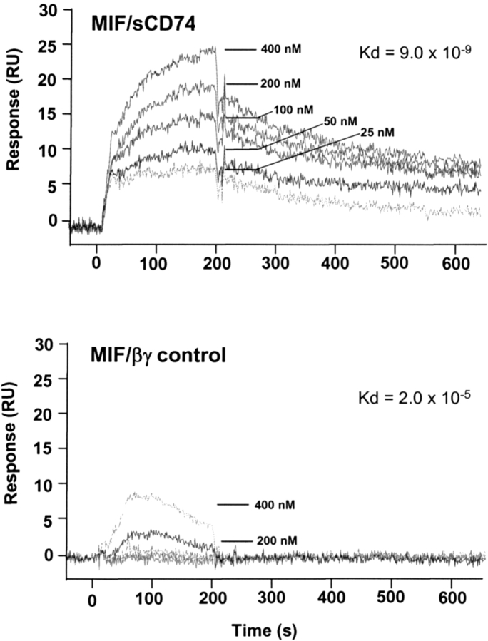
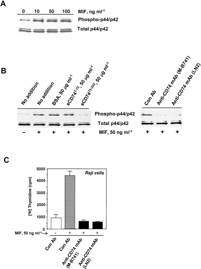
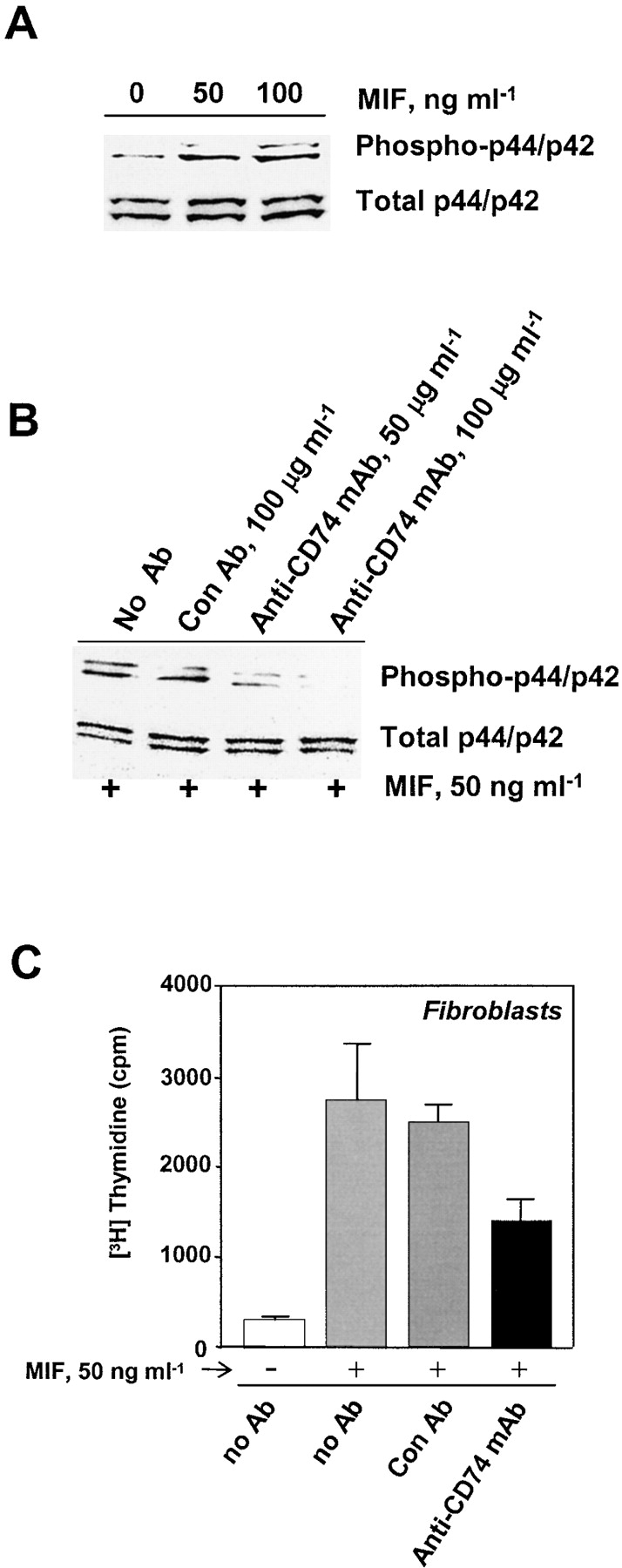
Macrophage migration inhibitory factor (MIF) accounts for one of the first cytokine activities to have been described, and it has emerged recently to be an important regulator of innate and adaptive immunity. MIF is an upstream activator of monocytes/macrophages, and it is centrally involved in the pathogenesis of septic shock, arthritis, and other inflammatory conditions. The protein is encoded by a unique but highly conserved gene, and X-ray crystallography studies have shown MIF to define a new protein fold and structural superfamily. Although recent work has begun to illuminate the signal transduction pathways activated by MIF, the nature of its membrane receptor has not been known. Using expression cloning and functional analysis, we report herein that CD74, a Type II transmembrane protein, is a high-affinity binding protein for MIF. MIF binds to the extracellular domain of CD74, and CD74 is required for MIF-induced activation of the extracellular signal-regulated kinase-1/2 MAP kinase cascade, cell proliferation, and PGE2 production. A recombinant, soluble form of CD74 binds MIF with a dissociation constant of approximately 9 x 10⁻⁹ Kd, as defined by surface plasmon resonance (BIAcore analysis), and soluble CD74 inhibits MIF-mediated extracellular signal-regulated kinase activation in defined cell systems. These data provide a molecular basis for MIF's interaction with target cells and identify it as a natural ligand for CD74, which has been implicated previously in signaling and accessory functions for immune cell activation.


Figures

Figure 1.
Figure 1.
Alexa-488–modified MIF (Alexa-MIF) shows retention of MIF biological activity in established assays. (A) Dose-dependent activation of the p44/p42 (ERK-1/2) MAP kinase cascade in IFN-γ–pretreated THP-1 monocytes (25). (B) Suppression of p53-dependent apoptosis in primary human fibroblasts (reference ; CM, complete medium; SFM, serum-free medium). MIF or Alexa-MIF was added at 50 ng/ml. Data shown are mean ± SD of triplicate wells and are representative of three independent experiments. (C) No difference in MIF's intrinsic tautomerase activity was observed in Alexa-MIF versus native (unconjugated) MIF using l-dopachrome methyl ester as a substrate (35).
Figure 2.
Figure 2.
Fluorescence analysis of Alexa-MIF binding to cells. (A) Flow cytometry analysis of the binding of Alexa-MIF to THP-1 monocytes. Competition for Alexa-MIF binding was performed in the presence of 20 μg/ml of unlabeled MIF. (B) Direct visualization of Alexa-MIF binding to THP-1 monocytes by confocal microscopy. THP-1 cells were incubated with 1 ng/ml IFN-γ for 72 h, and stained with Alexa-MIF (left) or with Alexa-MIF plus a 25-fold excess of unlabeled rMIF (middle). Cell-bound Alexa-MIF was rapidly internalized upon shifting cells from 4° to 37°C for 15 min (right). Magnification: 630.
Figure 3.
Figure 3.
Identification by expression cloning of CD74 as a cell surface binding protein for MIF. (A) Progressive enrichment by fluorescence-activated cell sorting of COS-7 cell transfectants showing MIF binding activity. (B) Structure of CD74 (35-kD isoform), and 3 of 10 representative CD74 cDNA clones with MIF binding activity. IC, TM, and EC are the intracellular, transmembrane, and extracellular domains, respectively. M1 and M17 refer to two sites of alternative translation initiation (42).
Figure 4.
Figure 4.
Fluorescence analysis of Alexa-MIF binding to CD74-expressing cells. (A) Flow cytometry analysis of the binding of Alexa-MIF to CD74-transfected versus control vector-transfected COS-7 cells (left), and binding of Alexa-MIF to CD74-transfected COS-7 cells incubated with anti-CD74 mAb (clone LN2) versus an isotypic mAb control (right, con Ab). The anti-CD74 mAb, LN2, is reactive with an epitope residing within 60 amino acids of the extracytoplasmic, COOH terminus of the protein (48). mAbs were added at 50 μg/ml, and the data shown are representative of at least three independent experiments. (B) Confocal microscopy images of a representative THP-1 cell (IFN-γ–pretreated) double stained with Alexa-MIF (left) and a rhodamine-labeled anti-CD74 mAb (middle). The merged images with yellow areas indicate colocalization of MIF and CD74 (right). The percent colocalization was calculated to be 69.2 ± 12.0 (P = 0.0308, n = 6 cells).
Figure 5.
Figure 5.
Biochemical evidence of MIF binding to CD74. (A) Cell-expressed CD74 binds MIF as detected by CD74 pull-down. Membrane-truncated CD74 (V5-CD741–72), full-length (V5-CD741–232), and NH2-terminal truncated CD74 (V5-CD7446–232) cDNAs were expressed in the pcDNA 3.1/V5-HisTOPO expression vector, transfected into COS-7 cells, and the protein products were precipitated by their expressed His tag. CD74 expression and recovery were monitored by Western blotting with anti-V5 (top). MIF was detected by Western blotting with anti-MIF (bottom). Vector: cells transfected with an empty vector control. (B) MIF binds to the extracellular domain of CD74 in vitro. 35S-CD74 protein was prepared in a coupled transcription and translation reaction using the different CD74 expression plasmids shown. Protein–protein interaction was assessed by measuring bound radioactivity in 96-well plates that were precoated with MIF (n = 6 wells per experiment). The data shown are representative of three experiments. (C) Soluble, extracellular domain CD74 (sCD7473–232) but not membrane-truncated CD74 (sCD741–72) inhibits MIF detection by sandwich ELISA. Increasing concentrations of MIF were captured by an immobilized anti-MIF mAb, followed by the addition of the sCD74 species shown and a biotinylated anti-MIF pAb (43). The bound complexes were detected with streptavidin-conjugated alkaline phosphatase and p-nitrophenylphosphate as a substrate.
Figure 6.
Figure 6.
High-affinity binding of MIF to CD74 measured by real-time, surface plasmon resonance (BIAcore analysis). Representative biosensorgrams of the interaction between sCD74 (sCD7473–232) and an MIF sensor chip as described in Materials and Methods (top). A control of MIF interaction with the membrane-associated G protein, βγ (bottom).
Figure 7.
Figure 7.
CD74 mediates MIF stimulation of p44/p42 (ERK-1/2) phosphorylation and PGE2 production in wild-type but not CD74-KO macrophages. Thioglycolate-elicited peritoneal macrophages were obtained from CD74+/+ and CD74−/− mice, and 6 × 10⁵ cells were stimulated with the indicated concentrations of MIF for 2.5 h. Cells were harvested, and the lysates were quantified for phospho-p44/p42 and total p44/p42 using specific antibodies as described in Materials and Methods. Supernatant PGE2 concentrations were measured by ELISA (10). Data shown are representative of three independent experiments.
Figure 8.
Figure 8.
CD74 mediates MIF stimulation of ERK-1/2 (p44/p42) phosphorylation and proliferation of human Raji B cells. (A) MIF initiates ERK-1/2 phosphorylation, and (B) sCD7473–232 and anti-CD74 mAb inhibit MIF-induced ERK-1/2 phosphorylation in Raji cells. Raji cells were stimulated with 50 ng/ml MIF for 2.5 h in the presence of an irrelevant protein (BSA), membrane-truncated CD74 (sCD741–72), extracellular domain CD74 (sCD7473–232), an isotype control antibody (Con Ab), or two anti-CD74 mAbs (clones M-B741 or LN2, each added at 50 μg/ml). Anti-CD63 mAb, which is directed to an irrelevant Raji cell surface protein (63), also did not block MIF-stimulated p44/p42 phosphorylation when compared with anti-CD74 mAb (not depicted). (C) Anti-CD74 mAb inhibits MIF-induced Raji cell proliferation. Raji cells were cultured as described in Materials and Methods, and stimulated with rMIF as shown in the presence of 50 μg/ml of the indicated antibodies. Anti-CD74 antibodies showed no effect on Raji cell proliferation in the absence of added MIF (not depicted).
Figure 9.
Figure 9.
CD74 mediates MIF stimulation of ERK-1/2 (p44/p42) phosphorylation and proliferation of CCL210 human lung fibroblasts. (A) MIF stimulates ERK-1/2 (p44/p42) phosphorylation, and (B) anti-CD74 mAb inhibits ERK-1/2 phosphorylation and proliferation of CCL210 human lung fibroblasts. Fibroblasts were stimulated with 50 ng/ml MIF for 2.5 h in the presence of an isotype control antibody (Con Ab) or the anti-CD74 mAb (clone LN2). (C) Anti-CD74 inhibits MIF-induced proliferation of human fibroblasts. Cells were stimulated for 2.5 h with 50 ng/ml rMIF in the presence of a Con Ab or anti-CD74 mAb (clone LN2), each at 100 μg/ml. Proliferation results are the mean ± SD of triplicate assays and are representative of at least three separate experiments. Anti-CD74 antibodies showed no effect on the proliferation of lung fibroblasts in the absence of added MIF (not depicted).

Similar articles

Cited by

References

    1. George, M., and J.H. Vaughn. 1962. In vitro cell migration as a model for delayed hypersensitivity. Proc. Soc. Exp. Biol. Med. 111:514–521. - PubMed
    1. David, J. 1966. Delayed hypersensitivity in vitro: its mediation by cell-free substances formed by lymphoid cell-antigen interaction. Proc. Natl. Acad. Sci. USA. 56:72–77. - PMC - PubMed
    1. Bloom, B.R., and B. Bennett. 1966. Mechanism of a reaction in vitro associated with delayed-type hypersensitivity. Science. 153:80–82. - PubMed
    1. Weiser, W.Y., P.A. Temple, J.S. Witek-Giannotti, H.G. Remold, S.C. Clark, and J.R. David. 1989. Molecular cloning of a cDNA encoding a human macrophage migration inhibitory factor. Proc. Natl. Acad. Sci. USA. 86:7522–7526. - PMC - PubMed
    1. Bernhagen, J., T. Calandra, R.A. Mitchell, S.B. Martin, K.J. Tracey, W. Voelter, K.R. Manogue, A. Cerami, and R. Bucala. 1993. MIF is a pituitary-derived cytokine that potentiates lethal endotoxaemia. Nature. 365:756–759. - PubMed

Publication types

MeSH terms

Substances