. 2003 Jun 2;197(11):1585-98.
doi: 10.1084/jem.20021868.

Accumulation of tissue factor into developing thrombi in vivo is dependent upon microparticle P-selectin glycoprotein ligand 1 and platelet P-selectin

Affiliations

Accumulation of tissue factor into developing thrombi in vivo is dependent upon microparticle P-selectin glycoprotein ligand 1 and platelet P-selectin

Shahrokh Falati et al. J Exp Med. .

Abstract

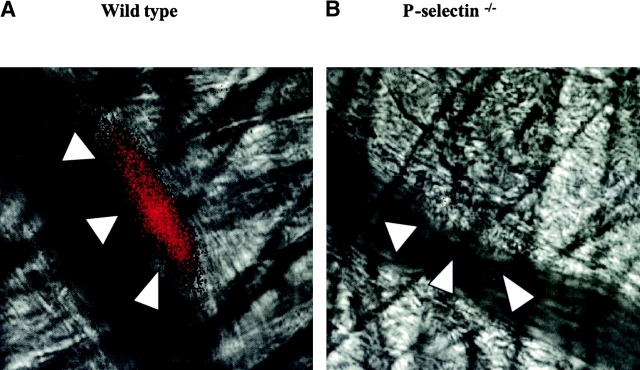
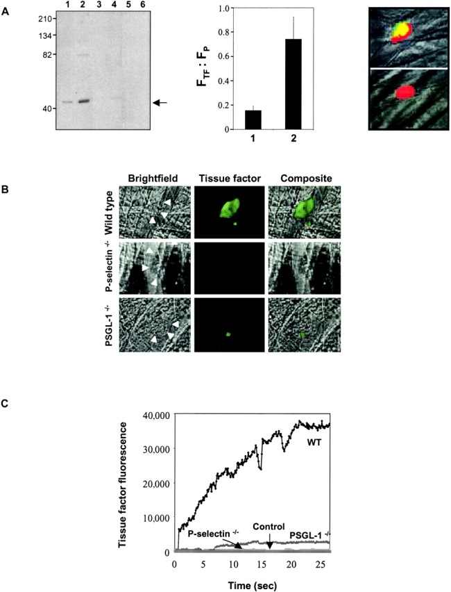
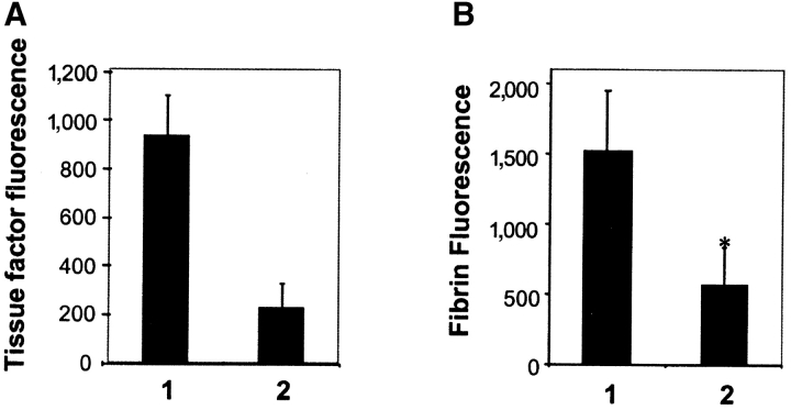
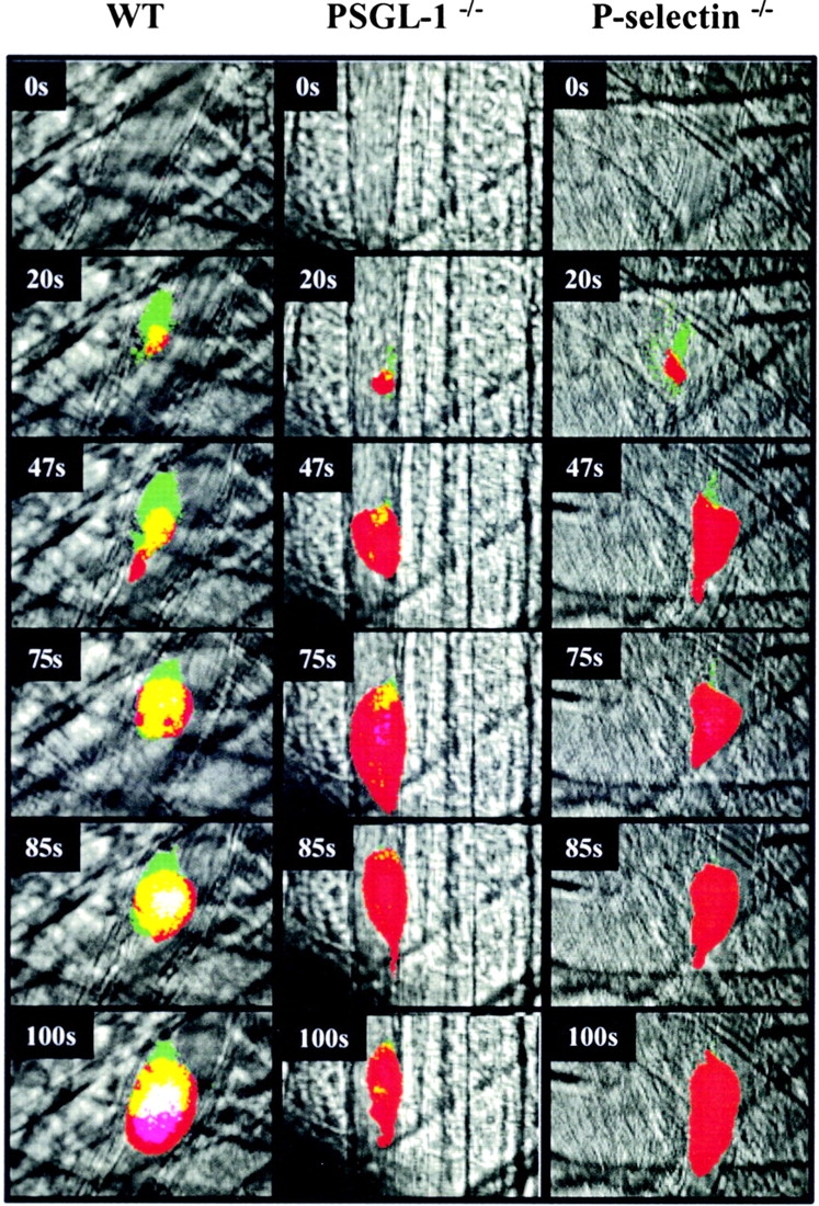
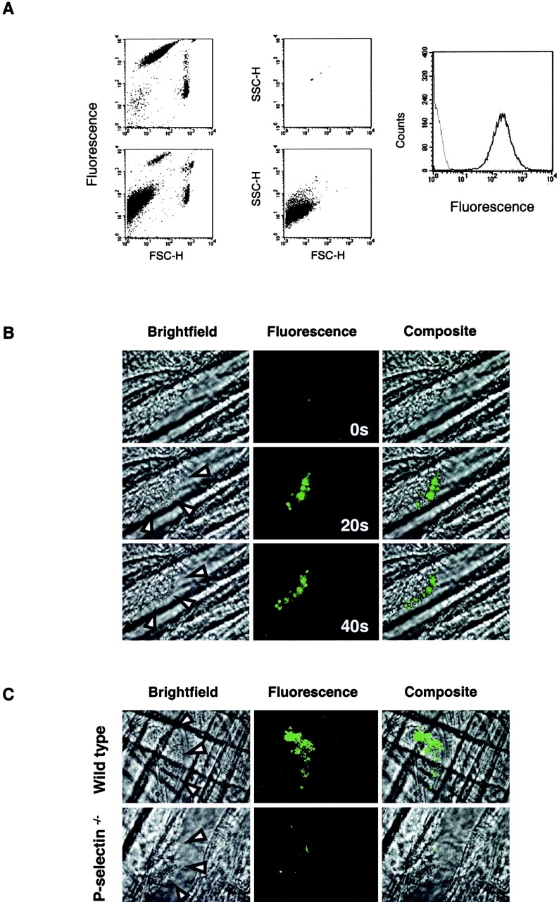
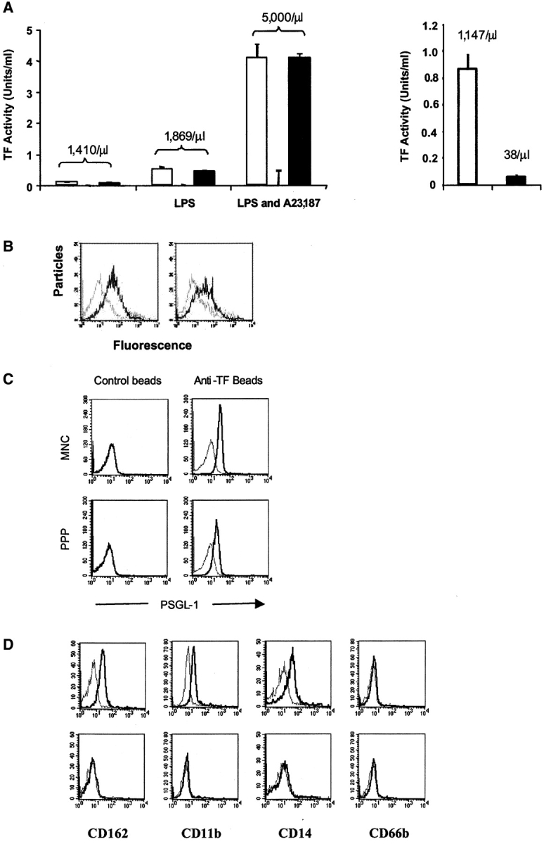
Using a laser-induced endothelial injury model, we examined thrombus formation in the microcirculation of wild-type and genetically altered mice by real-time in vivo microscopy to analyze this complex physiologic process in a system that includes the vessel wall, the presence of flowing blood, and the absence of anticoagulants. We observe P-selectin expression, tissue factor accumulation, and fibrin generation after platelet localization in the developing thrombus in arterioles of wild-type mice. However, mice lacking P-selectin glycoprotein ligand 1 (PSGL-1) or P-selectin, or wild-type mice infused with blocking P-selectin antibodies, developed platelet thrombi containing minimal tissue factor and fibrin. To explore the delivery of tissue factor into a developing thrombus, we identified monocyte-derived microparticles in human platelet-poor plasma that express tissue factor, PSGL-1, and CD14. Fluorescently labeled mouse microparticles infused into a recipient mouse localized within the developing thrombus, indicating that one pathway for the initiation of blood coagulation in vivo involves the accumulation of tissue factor- and PSGL-1-containing microparticles in the platelet thrombus expressing P-selectin. These monocyte-derived microparticles bind to activated platelets in an interaction mediated by platelet P-selectin and microparticle PSGL-1. We propose that PSGL-1 plays a role in blood coagulation in addition to its known role in leukocyte trafficking.


Figures

Figure 1.
Figure 1.
Expression of P-selectin in the developing thrombus. P-selectin was localized in the developing thrombus with Alexa 350–conjugated rat anti–mouse P-selectin antibody. The Alexa 350 fluorochrome image, collected digitally and presented as a red pseudocolor, is merged with the brightfield image. The images depicted are 160 s (wild-type mouse) and 200 s (P-selectin null mouse) after laser-induced endothelial injury. The boundaries of the thrombus are indicated by the arrowheads. A, wild-type mouse; B, P-selectin null mouse.
Figure 2.
Figure 2.
Tissue factor accumulation in the developing arterial thrombus. (A) Left: characterization of rabbit anti–mTF (152–166) antibodies. Western blot of tissue factor with affinity-purified rabbit anti–mTF (152–166) antibodies. STO cells, red cells, and platelets were lysed and the proteins in the lysate were separated on a 10% SDS-PAGE gel under reducing conditions. Lanes 1 and 5, 5 μg STO cell lysate; lanes 2 and 6, 7 μg STO cell lysate; lane 3, 5 μg murine red cell lysate; lane 4, 7 μg murine red cell lysate. Lanes 1–4 were developed with anti–mouse tissue factor antibody and lanes 5 and 6 were developed with an irrelevant antibody, anti-CD41. Arrow indicates molecular weight of tissue factor. Middle: comparison of tissue factor fluorescence detected with rabbit anti–mTF (152–166) antibodies (bar 2) or nonimmune IgG control (bar 1). The ratio of the integrated tissue factor fluorescence (FTF) to the integrated platelet fluorescence (FP) in thrombi generated in wild-type mice is shown based upon multiple independent experiments. P < 0.0042. Right: image of tissue factor in a thrombus detected using rabbit anti–mTF (152–166) antibodies. Top: Thrombus detected with anti-mTF (152–166) antibodies and anti-CD41 antibody directed against platelets. Bottom: Thrombus imaged with nonimmune IgG and anti-CD41 antibody directed against platelets. Red, platelets; green, tissue factor; yellow, platelets plus tissue factor. (B) Intravital images of arterial thrombi. Tissue factor was detected using Alexa 488–conjugated anti-tissue factor antibodies infused into the systemic circulation. The fluorescence image, recorded digitally, is presented as the green pseudocolor. Brightfield images of the thrombi are indicated by arrowheads. Images are representative of 12 thrombi formed in 2 arterioles in 2 mice of each genotype. (C) Time course of tissue factor antigen accumulation in the developing arterial thrombus in wild-type and genetically altered mice. Tissue factor was detected using sheep anti–tissue factor antibodies infused into the systemic circulation followed by Alexa 660–conjugated donkey anti–sheep IgG. Sheep IgG was substituted for anti-tissue factor antibody, control. Each curve represents the raw digital data of a single representative experiment. WT, wild-type mouse; PSGL−/−, PSGL-1 null mouse; P-selectin−/−, P-selectin null mouse.
Figure 3.
Figure 3.
Anti–P-selectin antibodies inhibit tissue factor and fibrin accumulation in the developing thrombus in wild-type mice. (A) Tissue factor in the thrombus formed was quantitated using Alexa 488–conjugated sheep anti–tissue factor antibodies from images derived by high speed widefield microscopy. Thrombi were formed in the absence of anti–P-selectin antibody in a wild-type mouse (bar 1). Subsequently, anti–P-selectin antibody (0.5 μg/g body weight) was infused and thrombi were formed in the presence of antibody (bar 2) between 5 and 30 min after antibody infusion. Data are at 60 s after injury and from seven thrombi formed in four arterioles before infusion of antibody and seven thrombi formed in four arterioles after infusion of antibody. Two mice were used. Mean ± SEM. (B) Experimental protocol was the same as described above except that fibrin was detected using Alexa 660–conjugated anti-fibrin–specific antibodies.
Figure 4.
Figure 4.
Fibrin deposition in the developing thrombus. (A) Left: Alexa 660–conjugated anti–mouse fibrin antibody and rat anti–mouse CD41 detected with Alexa 488–conjugated chicken anti–rat IgG were used to detect fibrin (red) and platelets (green). Overlap of the platelet and fibrin images is shown as a composite (yellow). When used, hirudin (1 U/g body weight) was infused immediately before initial thrombus formation. Fibrin and platelets were observed in thrombi formed 60 s after vessel injury. Images are representative of 10 thrombi formed in 6 arterioles in 3 mice of each genotype. Right: the ratio of the integrated fibrin fluorescence (FF) to the integrated platelet fluorescence (FP) in thrombi generated in wild-type mice, PSGL-1 null mice, and P-selectin null mice is shown (black) based upon multiple independent experiments. The fluorescence background in the absence of fibrin was determined in wild-type mice treated with hirudin. Error bars indicate standard error of the mean whereas * indicates statistical significance. (B) Images of the developing thrombus from 0–90 s. Platelets (green), fibrin (red), and platelet/fibrin composite (yellow). (C) Time course of fibrin formation in the developing thrombi of wild-type and genetically altered mice. Each curve represents the raw digital data of a single representative experiment. WT, wild-type mouse; PSGL−/−, PSGL-1 null mouse; P-selectin−/−, P-selectin null mouse.
Figure 5.
Figure 5.
Intravital imaging of platelet, tissue factor, and fibrin deposition in the developing thrombus of a wild-type mouse, PSGL-1 null mouse, or P-selectin null mouse after endothelial injury. Alexa 660–conjugated CD41 Fab fragments, Alexa 488–conjugated sheep anti–tissue factor antibodies, and Alexa 350–conjugated mouse anti–fibrin antibodies were infused into the systemic circulation. Representative composite images of the developing thrombus are shown. Red, platelets; green, tissue factor; blue, fibrin; yellow, platelets plus tissue factor; turquoise, tissue factor plus fibrin; magenta, platelets plus fibrin; white, platelets plus fibrin plus tissue factor. Fluorescence intensity in each channel was zeroed in images obtained before thrombus formation. Videos were continuously collected in four channels before and during laser injury and during thrombus development. To simplify analysis of the composite image, the dynamic range of the intensity of each pseudocolor was minimized. Images are representative of four thrombi formed in each of the mouse strains. Left, wild-type mouse; middle, PSGL-1 null mouse; right, P-selectin null mouse.
Figure 6.
Figure 6.
Calcein-labeled mouse microparticles accumulate into arterial thrombi. A leukocyte-rich blood cell preparation was labeled with calcein AM. (A) Flow cytometry. Two distinct populations are shown on the top left: a small, highly labeled cell population (platelets) and a larger population with lower calcein labeling (leukocytes). Upon the addition of A23187, a third population of calcein-labeled particles appeared (bottom left). 0.93 μm calibration microbeads were used to determine the limits of size of particles <1 μm (top middle). After the addition of A23187, the cells were removed by centrifugation, with >95% of the remaining particles in the supernatant <1 μm (bottom middle). These microparticles exhibited calcein fluorescence (black) compared with particles generated under the same conditions without the addition of calcein (gray; right). (B) Calcein-labeled microparticles were injected into the mouse circulation and their incorporation into arterial thrombi monitored over time by real-time widefield fluorescence videomicroscopy. Images are shown at 0, 20, and 40 s after thrombus formation. Left, brightfield image; arrow heads, the lumenal edge of the thrombus; middle, fluorescence image; right, composite brightfield and fluorescence images. Experiments are representative of three independent experiments. (C) Calcein-labeled microparticles derived from WEHI cells, a murine monocyte-like cell line, were infused into wild-type mice (top) or P-selectin null mice (bottom). The images were obtained 60 s after endothelial injury and the initiation of thrombus formation. Left, brightfield image; arrow heads, the lumenal edge of the thrombus; middle, fluorescence image; right, composite brightfield and fluorescence images.
Figure 7.
Figure 7.
Functional tissue factor and PSGL-1 on microparticles and presence of microparticles positive for tissue factor and PSGL-1. (A) Monocytes produce tissue factor–bearing microparticles. The medium from monocytes isolated and cultured in the absence of any additions, in the presence of LPS, or in the presence of LPS and A23187 was centrifuged for 30 s at 14,000 g to remove cells. The microparticles in the supernatant were counted and assayed for the presence of tissue factor activity. Left: open bars, tissue factor activity in the supernatant; gray bars, tissue factor activity after addition of anti-tissue factor antibody; black bars, tissue factor activity after the addition of nonimmune IgG. Right: supernatant was analyzed for the presence of microparticles before and after ultracentrifugation at 150,000 g for 120 min. Open bar, before ultracentrifugation; black bar, after centrifugation. The number of particles per microliter is indicated. (B) Microparticles express functional PSGL-1. P-selectin Ig chimera adsorbed to beads was incubated with calcein-labeled microparticles in the presence of buffer (black) or EDTA (gray; left) and anti–PSGL-1 antibody (gray) or isotype-matched control antibody (black; right). (C) Flow cytometric analysis of tissue factor–positive microparticles concentrated from human platelet–poor plasma (PPP; bottom) and mononuclear cell supernatant (MNC; top). These microparticles were incubated with beads containing tissue factor antibody (right) and beads containing nonimmune IgG (left). Black, fluorescently labeled anti–PSGL-1 antibody; gray, fluorescently labeled anti–PSGL-1 antibody plus 50-fold excess of unlabeled anti–PSGL-1 antibody. (D) Flow cytometric analysis of tissue factor–positive microparticles concentrated from human platelet null plasma. CD162 (PSGL-1), CD11b, CD14, CD66b. Top, beads coated with anti-tissue factor antibody; bottom, beads coated with nonimmune IgG; black, fluorescently labeled specific antibody; gray, fluorescently labeled specific antibody plus 50 fold excess of unlabeled specific antibody.

References

    1. McEver, R.P. 1994. Selectins. Curr. Opin. Immunol. 6:75–84. - PubMed
    1. Kansas, G.S. 1996. Selectins and their ligands: current concepts and controversies. Blood. 88:3259–3287. - PubMed
    1. Yang, J., B.C. Furie, and B. Furie. 1999. The biology of P-selectin glycoprotein ligand-1: its role as a selectin counterreceptor in leukocyte-endothelial and leukocyte-platelet interaction. Thromb. Haemost. 81:1–7. - PubMed
    1. Hsu-Lin, S., C.L. Berman, B.C. Furie, D. August, and B. Furie. 1984. A platelet membrane protein expressed during platelet activation and secretion. Studies using a monoclonal antibody specific for thrombin-activated platelets. J. Biol. Chem. 259:9121–9126. - PubMed
    1. McEver, R.P., and M.N. Martin. 1984. A monoclonal antibody to a membrane glycoprotein binds only to activated platelets. J. Biol. Chem. 259:9799–9804. - PubMed

Publication types