. 2003 Jun;15(6):1375-85.
doi: 10.1105/tpc.010967.

PmSUC3: characterization of a SUT2/SUC3-type sucrose transporter from Plantago major

Affiliations

PmSUC3: characterization of a SUT2/SUC3-type sucrose transporter from Plantago major

Inga Barth et al. Plant Cell. 2003 Jun.

Abstract

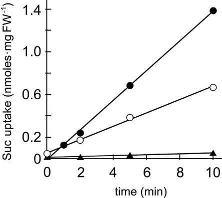
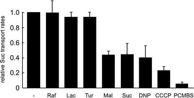
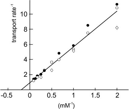
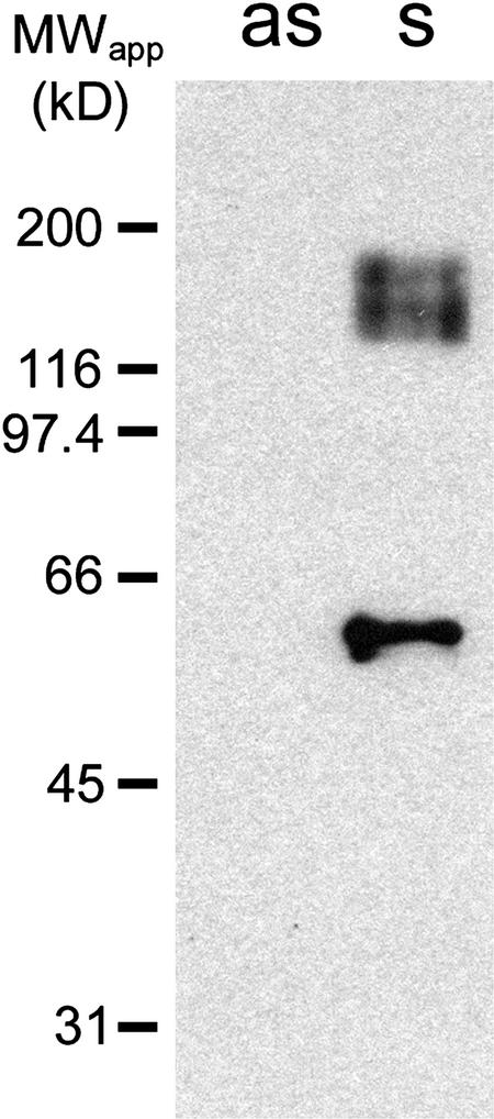
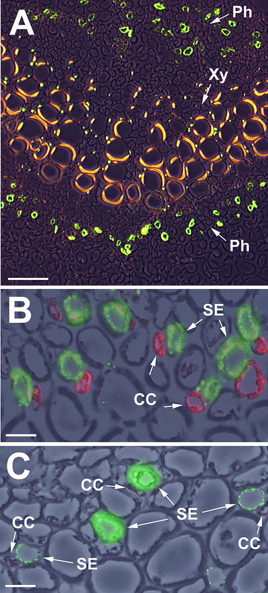
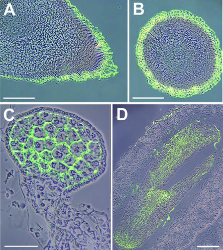
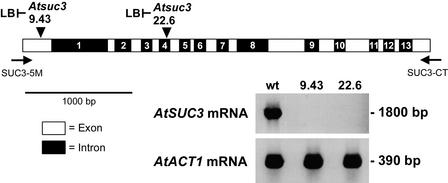
Higher plants possess medium-sized gene families that encode plasma membrane-localized sucrose transporters. For several plant species, it has been shown that at least one of these genes (e.g., AtSUC3 in Arabidopsis and LeSUT2 in tomato) differs from all other family members in several features, such as the length of the open reading frame, the number of introns, and the codon usage bias. For these reasons, and because two of these proteins did not rescue a yeast mutant defective in sucrose utilization, it had been speculated that this subgroup of transporters might have sensor functions. Here, we describe the detailed functional characterization and cellular localization of PmSUC3, the orthologous transporter from the Plantago major transporter family. The PmSUC3 protein is localized in the sieve elements of the Plantago phloem and mediates the energy-dependent transport of sucrose and maltose. In contrast to the situation in solanaceous plants, PmSUC3 is not colocalized with PmSUC2, the source-specific, phloem-loading sucrose transporter of Plantago. Moreover, PmSUC3 also was identified in sieve elements of sink leaves and in several nonphloem cells and tissues. Arguments for and against a potential sensor function for this type of sucrose transporter are presented, and the role of this type of transporter in the regulation of sucrose fluxes is discussed.


Figures

Figure 1.
Figure 1.
Comparison of the Deduced Protein Sequences of PmSUC3 and the Homologous AtSUC3 Protein from Arabidopsis. Sequences were aligned with the program SeqVu (James Gardner, Garvan Institute of Medical Research, Sydney, Australia), and residues identical in both sequences are highlighted. Regions corresponding to the N-terminal and central extensions in SUT2/SUC3-type proteins are underlined.
Figure 2.
Figure 2.
PmSUC3 Can Be Expressed in Yeast Cells and Catalyzes the Uptake of 14C-Sucrose. Transport rates for 14C-sucrose were determined with the transgenic yeast cells expressing PmSUC3 in the sense (open circles; strain IBY20s) or the antisense (closed triangles; strain IBY20as) orientation. The initial concentration of 14C-sucrose was 1 mM in all experiments. The addition of d-glucose (closed circles; final concentration of 10 mM) clearly enhances the transport rates of PmSUC3. FW, fresh weight.
Figure 3.
Figure 3.
Transport Properties of PmSUC3 in Yeast Cells. Transport of 14C-sucrose (1 mM) by strain IBY20s was analyzed in the presence of uncouplers (DNP or CCCP) or in the presence of the sulfhydryl group inhibitor PCMBS. Inhibitors were added to a final concentration of 50 μM. Transport of 14C-sucrose also was analyzed in the presence of other potential substrates added at a 10-fold excess (final concentrations of 10 mM). Each bar represents results from at least three independent analyses. Lac, lactose; Mal, maltose; Raf, raffinose; Suc, sucrose; Tur, turanose.
Figure 4.
Figure 4.
Km Value of PmSUC3 for Sucrose Transport in Transgenic Yeast Cells. The Lineweaver-Burk diagram shown contains data from three independent experiments represented by three different symbols. From the individual values of the three analyses, a Km value of 5.5 ± 1.1 mM was calculated.
Figure 5.
Figure 5.
Identification of PmSUC3 in Transgenic Yeast Cells. SDS-solubilized plasma membrane proteins (4 μg/lane) from yeast cells expressing PmSUC3 in the sense (s = IBY20s) or the antisense (as = IBY20as) orientation were separated on a polyacrylamide gel, transferred to a nitrocellulose filter, and incubated with anti-PmSUC3 antiserum at a dilution of 1:500. Binding of antibodies to PmSUC3 was detected with anti-rabbit IgG antiserum conjugated to peroxidase. MWapp, apparent molecular mass.
Figure 6.
Figure 6.
Immunolocalization of PmSUC3 in Sieve Elements of the Plantago Phloem. (A) Cross-section through the vascular bundle of a Plantago leaf treated with anti-PmSUC3/fluorescein isothiocyanate–conjugated anti-rabbit antiserum (green fluorescence in the phloem [Ph]). A photograph taken under white light and a photograph taken under excitation light were superimposed. The yellow fluorescence of xylem vessels in the center (Xy) results from phenolic compounds in the walls of these cells. Bar = 50 μm. (B) Cross-section through a medium-sized vascular bundle of a Plantago source leaf double stained with anti-PmSUC3/Alexa Fluor 488 goat anti-rabbit IgG (green fluorescence) and anti-PmSUC2/Alexa Fluor 546 goat anti-mouse IgG (red fluorescence). Three photographs (one taken under white light and two taken under excitation light) were superimposed. CC, companion cell; SE, sieve element. Bar = 1 μm. (C) Cross-section through a medium-sized vascular bundle of a Plantago sink leaf double stained with anti-PmSUC3/Alexa Fluor 488 goat anti-rabbit IgG (green fluorescence) and anti-PmSUC2/Alexa Fluor 546 goat anti-mouse IgG (red fluorescence, which was not detectable in sinks). Three photographs (one taken under white light and two taken under excitation light) were superimposed. Bar = 1 μm.
Figure 7.
Figure 7.
Identification of PmSUC3 in the Root Tips of Plantago. (A) Longitudinal section through a Plantago root tip. Two photographs were superposed, one taken under white light and one taken under excitation light to visualize Alexa Fluor 488–decorated PmSUC3. (B) Cross-section through a Plantago root tip presented as described for (A). (C) Longitudinal section through a Plantago embryo in the globular state presented as described for (A). (D) Longitudinal section through a Plantago embryo in the torpedo state presented as described for (A). Bars = 50 μm for (C) and 100 μm for (A), (B), and (D).
Figure 8.
Figure 8.
Identification of T-DNA Insertions in the Arabidopsis AtSUC3 Gene. The positions of the T-DNA insertions in the Arabidopsis lines Atsuc3-9.43 (first exon) and Atsuc3-22.6 (fourth intron) are indicated by arrowheads, and binding sites for the PCR primers SUC3-5M and SUC3-CT are represented by arrows. The orientations of the identified left borders (LB) of the T-DNA insertions are indicated. The indicated primers were used to perform RT-PCR on total RNA isolated from Arabidopsis wild-type (wt) plants or from the two different T-DNA insertion lines. RT-PCR products were identified only with RNA from wild-type plants. In control reactions, fragments of the Arabidopsis ACT1 mRNA (An et al., 1996) were amplified from all RNA preparations.

References

    1. An, Y.Q., Huang, S., McDowell, J.M., McKinney, E.C., and Meagher, R.B. (1996). Conserved expression of the Arabidopsis ACT1 and ACT3 actin subclass in organ primordia and mature pollen. Plant Cell 8, 15–30. - PMC - PubMed
    1. Anderson, J.A., Huprikar, S.S., Kochian, L.V., Lucas, W.J., and Gaber, R.F. (1992). Functional expression of a probable Arabidopsis thaliana potassium channel in Saccharomyces cerevisiae. Proc. Natl. Acad. Sci. USA 89, 3736–3740. - PMC - PubMed
    1. Aoki, N., Hirose, T., Scofield, G.N., Whitfeld, P.R., and Furbank, R.T. (2003). The sucrose transporter gene family in rice. Plant Cell Physiol. 44, 223–232. - PubMed
    1. Arabidopsis Genome Initiative (2000). Analysis of the genome sequence of the flowering plant Arabidopsis thaliana. Nature 408, 796–815. - PubMed
    1. Barker, L., Kühn, C., Weise, A., Schulz, A., Gebhardt, C., Hirner, B., Hellmann, H., Schulze, W., Ward, J.M., and Frommer, W.B. (2000). SUT2, a putative sucrose sensor in sieve elements. Plant Cell 12, 1153–1164. - PMC - PubMed

Publication types

MeSH terms

Associated data