

Comment
doi: 10.1073/pnas.1332695100.
Epub 2003 Jun 2.
Dengue virus envelope glycoprotein structure: new insight into its interactions during viral entry
Affiliations
- PMID: 12782795
- PMCID: PMC165800
- DOI: 10.1073/pnas.1332695100
Item in Clipboard
Comment
Dengue virus envelope glycoprotein structure: new insight into its interactions during viral entry
Proc Natl Acad Sci U S A.
.
No abstract available
Figures
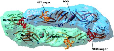
Semitransparent surface representation of the dengue virus E protein. The two subunits in the dimer are shown in different shades of blue. The backbone of the molecule is superposed in a yellow ribbon representation. The two glycosylation sites are indicated. To illustrate the location and size of the sugar moiety on a molecule produced in insect cells, the complete high-mannose glycans have been modeled, superposed to the core N-acetyl glucosamine residues that were visible in the crystal structure. The sugars linked to Asn-67 and -153 are shown as yellow and red sticks, respectively. The β-octyl glucoside molecule (indicated by bOG) is displayed as spheres colored according to atom type (red, oxygen; gray, carbon). It lies in a pocket at the hinge region between domains I and II. Figure prepared with the program ribbons (19).
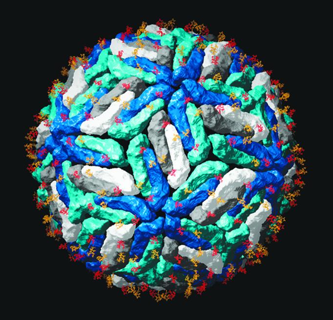
Carbohydrate distribution on the viral surface. The dengue virus E protein was superposed on the Protein Data Bank ID code 1K4R coordinates, which correspond to the tick-borne encephalitis E protein ectodomain placed in the reconstruction of dengue virus obtained by cryoelectron microscopy (18). Each dengue E dimer is represented as in Fig. 1, with the dimers that lie at the icosahedral twofold axis in dark and light gray, and the dimers lying on local twofold axes in two shades of blue. The dark- and light-blue subunits form the five- and threefold contacts, respectively. The sugars are as indicated in Fig. 1, with the same color coding. The high-mannose glycans present at the surface of mosquito-grown virions have been identified as important for the interactions with DC-SIGN in the initial steps leading to entry. Figure prepared with the program ribbons (19).
Comment on
-
A ligand-binding pocket in the dengue virus envelope glycoprotein.Proc Natl Acad Sci U S A. 2003 Jun 10;100(12):6986-91. doi: 10.1073/pnas.0832193100. Epub 2003 May 20. Proc Natl Acad Sci U S A. 2003. PMID: 12759475 Free PMC article.
References
Publication types
MeSH terms
Substances
LinkOut - more resources
Full Text Sources
Other Literature Sources
