. 2003 Jul;4(7):723-8.
doi: 10.1038/sj.embor.embor866.

Dendritic-cell-specific ICAM3-grabbing non-integrin is essential for the productive infection of human dendritic cells by mosquito-cell-derived dengue viruses

Affiliations

Dendritic-cell-specific ICAM3-grabbing non-integrin is essential for the productive infection of human dendritic cells by mosquito-cell-derived dengue viruses

Erika Navarro-Sanchez et al. EMBO Rep. 2003 Jul.

Abstract

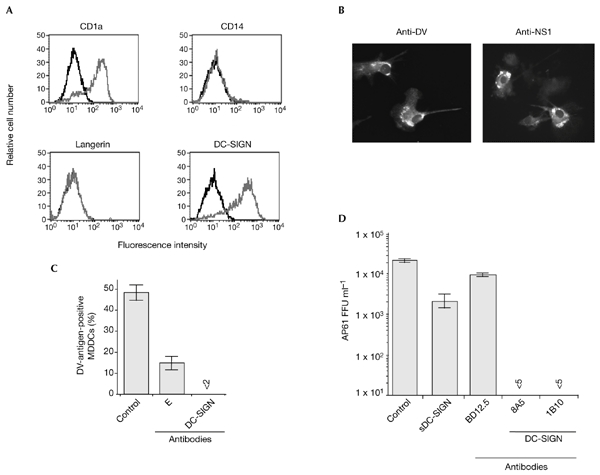
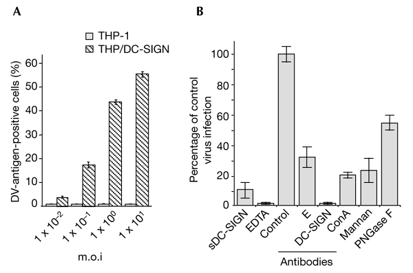
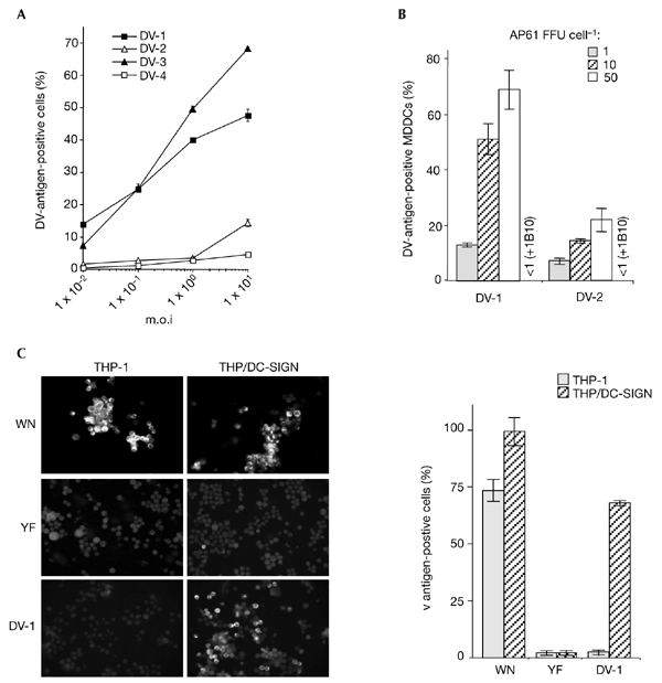
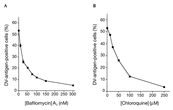
Dengue virus (DV) is a mosquito-borne flavivirus that causes haemorrhagic fever in humans. DV primarily targets immature dendritic cells (DCs) after a bite by an infected mosquito vector. Here, we analysed the interactions between DV and human-monocyte-derived DCs at the level of virus entry. We show that the DC-specific ICAM3-grabbing non-integrin (DC-SIGN) molecule, a cell-surface, mannose-specific, C-type lectin, binds mosquito-cell-derived DVs and allows viral replication. Conclusive evidence for the involvement of DC-SIGN in DV infection was obtained by the inhibition of viral infection by anti-DC-SIGN antibodies and by the soluble tetrameric ectodomain of DC-SIGN. Our data show that DC-SIGN functions as a DV-binding lectin by interacting with the DV envelope glycoprotein. Mosquito-cell-derived DVs may have differential infectivity for DC-SIGN-expressing cells. We suggest that the differential use of DC-SIGN by viral envelope glycoproteins may account for the immunopathogenesis of DVs.


Figures

Figure 1
Figure 1
Anti-DC-SIGN antibodies and soluble DC-SIGN inhibit the ability of dengue virus type 1 to infect DC-SIGN-expressing monocyte-derived dendritic cells. (A) The expression levels of the cell-surface markers CD1a, CD14, langerin and DC-SIGN on immature human monocyte-derived dendritic cells (MDDCs) were assessed by direct fluorescence. DC-SIGN-expressing cells were detected using the anti-DC-SIGN monoclonal antibody, 1B10. The relative fluorescence intensity was measured by FACScan analysis and the histograms show the binding of the specific antibody (grey) and isotype-matched control antibody (black). (B) MDDCs were infected for 40 h with dengue virus (DV)-1 at a multiplicity of infection of 5 and were immunostained with anti-DV-1-specific hyperimmune mouse ascites fluids (HMAF) or anti-DV-1 NS1 monoclonal antibody (anti-NS1), and were observed using a fluorescent microscope. (C) Before infection, MDDCs were mock-treated (control) or incubated with 20 µg ml−1 monoclonal antibody 1B10 (DC-SIGN) or a 1:50 dilution of anti-DV E monoclonal antibody 9D12 (E) in RPMI, 0.2% BSA, for 20 min at 25 °C. Treated MDDCs were infected for 2 h at 37 °C in the continuous presence of antibodies. DV antigens were visualized by an immunofluorescence assay using anti-DV-1-specific HMAF. (D) Before infection, MDDCs were mock-treated (control) or were incubated with 20 µg ml−1 anti-LCMV (lymphocytic choriomeningitis virus) monoclonal antibody (BD12.5), anti-DC-SIGN monoclonal antibody (8A5) or anti-DC-SIGN monoclonal antibody (1B10). FGA/NA d1d virus (1 × 10⁵ AP61 focus-forming units (FFU)) was mixed with 10 µg ml−1 sDC-SIGN in RPMI, 0.2% BSA, for 20 min at 25 °C. MDDCs were infected for 2 h at 37 °C in the continuous presence of inhibitors. Infectious virus particles produced in the supernatants were titred. Each experimental point represents the mean ± s.d. of results obtained from three chambers. Magnification in (B), ×100. DC-SIGN, dendritic-cell-specific ICAM3-grabbing non-integrin; sDC-SIGN, the soluble, tetrameric ectodomain of DC-SIGN.
Figure 2
Figure 2
The lectin DC-SIGN is crucial for the entry of dengue virus into monocytic cells. THP-1 and TPH/DC-SIGN cells infected with dengue virus (DV)-1 FGA/NA d1d were analysed for viral protein production as described in the legend for Fig. 1. (A) Cells infected at various multiplicities of infection (m.o.i.s) were analysed using an immunofluorescence assay. (B) THP/DCsIGN cells were infected with FGA/NA d1d virus at m.o.i.s of 5 (columns labelled EDTA and antibodies) or 1 (columns labelled sDC-SIGN, mannan and ConA). Before infection, THP/DC-SIGN cells were incubated with either 20 µg ml−1 mannan, 5 mM EDTA, 20 µg ml−1 monoclonal antibody BD12.5 (control), a 1:50 dilution of monoclonal antibody 9D12 (E), or 20 µg ml−1 monoclonal antibody 1B10 (against DC-SIGN) as described in the legend to Fig. 1. FGA/NA d1d virus (1 × 10⁵ AP61 focus-forming units) was mixed with 10 µg ml−1 sDCsIGN (20 min at 25 °C) or incubated with 25 µg ml−1 ConA (1 h at 37 °C) or N-glycosidase F (PGNase F; see the Methods section). Cells positive for DV antigens are expressed as a percentage of untreated, DV-infected THP/DC-SIGN cells (percentage of control virus infection). ConA, concanavalin A; DC-SIGN, dendritic-cell-specific ICAM 3-grabbing non-integrin; sDC-SIGN, the soluble, tetrameric ectodomain of DC-SIGN.
Figure 3
Figure 3
Flavivirus infectivity in DC-SIGN-expressing monocytic cells. Cells infected with flaviviruses were assayed 25 h (monocyte-derived dendritic cells (MDDCs)) or 40 h (THP/DC-SIGN) post-infection. for the presence of viral antigens, using the immunofluorescence assay described in the legend of Fig. 1. (A) THP/DCsIGN cells were infected with strain FGA/NA d1d of dengue virus (DV)-1, the Jam strain of DV-2, strain H-87 of DV-3 or strain H-241 of DV-4, at various multiplicities of infection (m.o.i.s) (B) MDDCs were infected with highly purified DV-1 FGA/NA d1d and DV-2 Jam viruses at various m.o.i.s. Before infection, MDDCs infected at an m.o.i. of 10 were incubated with 20 mg ml−1 monoclonal antibody 1B10 (+1B10), as described in the legend for Fig. 1. Experiments were performed twice using two MDCC donors. The results of one representative experiment are shown. (C) THP-1 and THP/DCsIGN cells were infected with strain IS-98-ST1 of West Nile (WN) virus (m.o.i. of 5), vaccine strain 17D of yellow fever (YF) virus (m.o.i. of 50) or DV-1 (m.o.i. of 5) and viral antigens were visualized by immunofluorescence assays with specific hyperimmune mouse ascites fluids. Each experimental point represents the mean ± s.d. of results obtained from three chambers. Magnification in left panel of (C), x100. DC-SIGN, dendritic-cell-specific ICAM3-grabbing non-integrin.
Figure 4
Figure 4
DC-SIGN-mediated dengue virus entry requires acidification. THP/DC-SIGN cells were infected with dengue virus (DV)-1 FGA/NA d1d (multiplicity of infection of 1) in the presence of increasing doses of bafilomycin A1 (A) or chloroquine (B), as described in the Methods section. Treated cells were assayed 40 h post-infection for the presence of viral antigens using an immunofluorescence assay, as described in the legend for Fig. 1. Each data point represents the mean ± s.d. of results from two chambers. DCsIGN, dendritic-cell-specific ICAM3-grabbing non-integrin.

References

    1. Alvarez C.P., Lasala F., Carillo J., Muniz O., Corbi A.L. & Delgado R. (2002) C-type lectins DCsIGN and L-SIGN mediate cellular entry by Ebola virus in cis and in trans. J.Virol., 76, 6841–6844. - PMC - PubMed
    1. Bielefeldt-Ohmann H., Meyer M., Fitzpatrick D.R. & Mackenzie J.S. (2001) Dengue virus binding to human leukocyte cell lines: receptor usage differs between cell types and virus strains. Virus Res., 73, 81–89. - PubMed
    1. Chen Y., Maguire T., Hileman R.E., Fromm J.R., Esko J.D., Linhardt R.J. & Marks R.M. (1997) Dengue virus infectivity depends on envelope protein binding to target cell heparane sulfate. Nature Med., 3, 866–871. - PubMed
    1. Courageot M.-P., Frenkiel M.-P., Duarte Dos Santos C., Deubel V. & Desprès P. (2000) α-Glucosidase inhibitors reduce dengue virus production by affecting the initial steps of virion morphogenesis in the endoplasmic reticulum. J. Virol., 74, 564–572. - PMC - PubMed
    1. Desprès P., Girard M. & Bouloy M. (1991) Characterization of yellow fever virus E and NS1 proteins expressed in Vero and Spodoptera frugiperda cells. J. Gen. Virol., 72, 1331–1342. - PubMed

Publication types

MeSH terms