Comparative Study
. 2003 Aug;52(8):497-505.
doi: 10.1007/s00262-003-0377-8. Epub 2003 Jun 3.

Recognition of PSA-derived peptide antigens by T cells from prostate cancer patients without any prior stimulation

Affiliations
Comparative Study

Recognition of PSA-derived peptide antigens by T cells from prostate cancer patients without any prior stimulation

Nitya G Chakraborty et al. Cancer Immunol Immunother. 2003 Aug.

Abstract

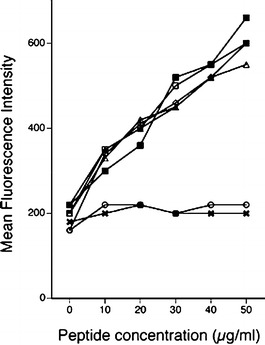
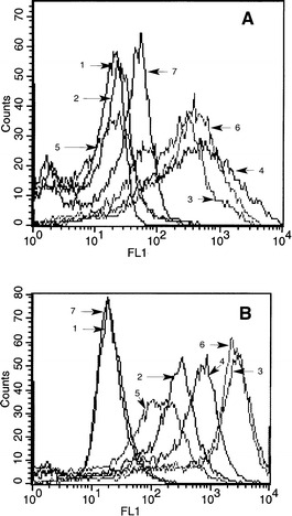
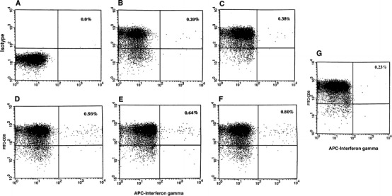
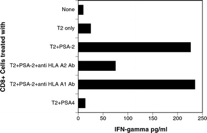
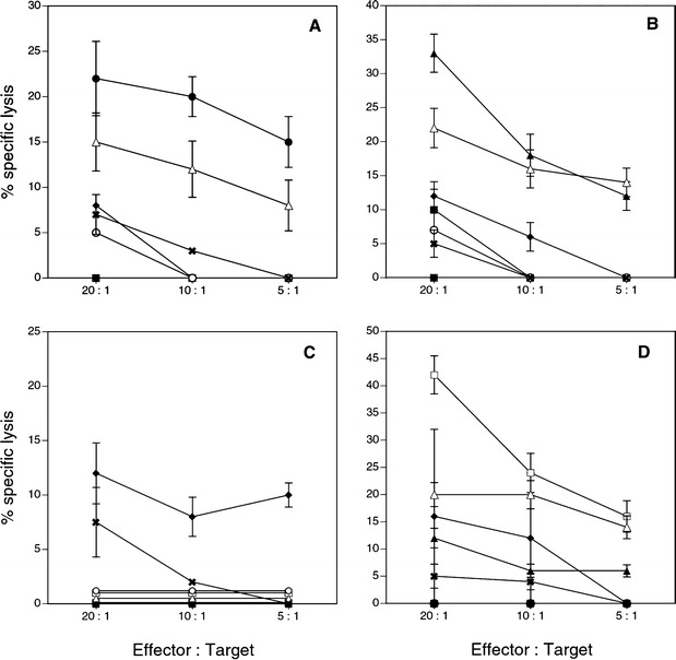
Prostate-specific antigen (PSA) is a valuable marker antigen for prostate cancer. Lately considerable interest has been generated in the prospect of developing a vaccine for prostate cancer with PSA-derived peptide epitopes to induce cytotoxic T-cell (CTL) response. We report here that T cells capable of exhibiting PSA epitope-specific effector function-in their native state, i.e, without having to be further stimulated, in vitro-are detectable in more than half of the prostate cancer patients we studied. Ex vivo cultured autologous dendritic cells (DC) were used to present four HLA-A2-binding PSA peptide epitopes to freshly isolated peripheral blood lymphocytes (PBL) from patients and healthy volunteers. Ten out of 14 patients' PBL recognized at least one of the four peptides and 6 out of 10 patients' PBL recognized more than one peptide antigen as measured by IFN-gamma secretion upon stimulation of the PBL with the peptide antigen. Intracytoplasmic cytokine analysis for IFN-gamma in purified CD8(+) cells after stimulation with peptide antigens was tested in 6 patients and this technique demonstrated a similar response. Freshly isolated and purified CD8(+) cells when tested, also recognized the epitopes, as measured by IFN assay, when presented by transporter associated with antigen-processing (TAP) deficient T2 cells in an MHC-I restricted fashion. PBL from 9 normal donors when tested in identical fashion did not show any IFN-gamma production in recognition to the peptide antigens. Interestingly, neither of these CD8(+) T cells having IFN-gamma-producing ability did show any cytolytic activity in their native state against peptide loaded target cells or tumor cells when tested in cytotoxicity assay. In long term cocultures stimulation of purified CD8(+) T cells with matured DC pulsed with PSA peptides generated a PSA-specific CTL response in 4 of 6 patients studied and in 2 of 9 normal donors. While our observations of CTL generation are consistent with the prior reports that have demonstrated that specific CD8(+) CTL could be generated which recognize PSA-derived epitopes by in vitro stimulation by one means or another, this observation that IFN-gamma-producing CD8(+) T cells are present in patients which are antigen experienced, and do not require in vitro stimulation, is novel and has major implications for prostate cancer vaccine preparation.


Figures

Fig. 1.
Fig. 1.
Binding of PSA-derived peptides to HLA-A2 on T2 A2 cells. Peptides were analyzed for binding to T2 A2 cells as described. This describes the binding of peptides to A2 when different concentrations (10–50 μg/ml) of peptides were used. X T2 cells without any peptide, open circle T2 + MAGE-1, open square T2 + PSA-1, solid square T2 + PSA-2, open triangle T2 + PSA-3, solid triangle T2 + PSA-4, open diamond T2 + MART-1
Fig. 2A, B.
Fig. 2A, B.
Phenotypic analysis of dendritic cells. A Different surface markers on DC after 7 days in culture in presence of GM-CSF and IL-4, immature DC. B Surface markers on DC further matured with LPS + IFN-γ for additional 24 hr, matured DC. 1 Isotypecontrol, 2 CD80, 3 CD86, 4 MHC-I, 5 CD83, 6 CD40, and 7 CD14. MFI of various markers on immature DC as in A are Isotype = 8.3, CD80 = 17.0, CD86 = 289.1, MHC-I = 139.0, CD83 = 9.2, CD40 = 232.5, and CD14 = 21.7. MFI of various markers on matured DC in 2B are Isotype = 14.2, CD80 = 148.7, CD86 = 1118, MHC-1 = 356.9, CD83 = 71.2, CD40 = 1137.6, and CD14 = 15.9.
Fig. 3A–G.
Fig. 3A–G.
Intracellular cytokine analysis. CD8+ cells from PBL were isolated by positive selection using Dynal magnetic beads. Cells were stimulated with PSA-peptide-pulsed autologous DC for 6 h. Cells were stained with anti-CD8 antibody conjugated with FITC (Y axis) and then stained for intracellular staining with antihuman IFN-γ conjugated with APC (X axis) according to the method described. A Isotype matched control. B Cells stimulated with DC only. C Cells stimulated with PSA-1 peptide-pulsed DC. D Cells stimulated with PSA-2 pulsed DC. E Cells stimulated with PSA-3-pulsed DC. F Cells stimulated with PSA-4-pulsed DC. G Cells stimulated with Mart-1 peptide-pulsed DC
Fig. 4.
Fig. 4.
Response to PSA peptides presented by T2 cells. Purified CD8+ cells from a patient (PD) who was responding against PSA-2 were stimulated with T2 cells pulsed with PSA-2 in presence and absence of anti-HLA-A2 antibody (1 μg/ml). PSA-4 peptide was used as control for the experiment
Fig. 5A–D.
Fig. 5A–D.
Cytotoxicity assay with in vitro expanded peptide-specific CTL. CD8+ cells were stimulated in vitro with matured autologous DC pulsed with one of the PSA-derived peptides. Cytotoxicity was measured by 51Cr release from labeled different target cells. Target cells were T2 cells alone, T2 cells pulsed with one of the four PSA peptides, T2 cells pulsed with Flu peptide, HLA-A2 positive and PSA positive prostate cancer cell line LN CaP, and HLA-A2 positive and PSA negative melanoma cell line CS-M. A CTL activity with responding CD8+ cells stimulated with DC pulsed with PSA-1. B CTL activity with responding CD8+ cells stimulated with DC pulsed with PSA-2. C CTL activity with responding CD8+ cells stimulated with DC pulsed with PSA-3. D CTL activity with responding CD8+ cells stimulated with DC pulsed with PSA-4. Solid square % specific lysis of the target T2 cells alone. Solid circle % specific lysis of the target T2 cells pulsed with peptide PSA-1. Solid triangle % specific lysis of the target T2 cells pulsed with peptide PSA-2. Solid diamond % specific of the target T2 cells pulsed with peptide PSA-3. Open square % specific lysis of the target T2cells pulsed with peptide PSA-4. Open circle % specific lysis of the target T2 cells pulsed with Flu peptide. Open triangle % specific lysis of the target LN CaP tumor cells. X % specific lysis of HLA A2 positive melanoma tumor target CS-M cells

Similar articles

Cited by

References

    1. Alexander Urology. 1998;51:150. doi: 10.1016/S0090-4295(97)00480-9. - DOI
    1. Altman Science. 1996;274:94. - PubMed
    1. Anderson J Immunol. 1993;151:3407. - PubMed
    1. Baecher-Allan J Immunol. 2001;167:1245. - PubMed
    1. Boon J Exp Med. 1996;183:725. - PMC - PubMed

Publication types

MeSH terms