

Evaluation of a transpedicular drill guide for pedicle screw placement in the thoracic spine
- PMID: 12783286
- PMCID: PMC3468009
- DOI: 10.1007/s00586-003-0549-4
Evaluation of a transpedicular drill guide for pedicle screw placement in the thoracic spine
Abstract
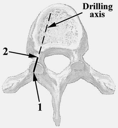
Insertion of pedicle screws in the thoracic spine is technically difficult and may lead to major complications. Although many computer-assisted systems have been developed to optimize pedicle screw insertion, these systems are expensive, not user-friendly and involve significant radiation from pre-operative computed tomographic (CT) scan imaging. This study describes and evaluates a transpedicular drill guide (TDG) designed to assist in the proper placement of pedicle screws in the thoracic spine. Pilot holes were made manually using the TDG in the thoracic spine (T1-T11) of three human cadavers before inserting 4.5-mm-diameter screws. CT scans followed by visual inspection of the spines were performed to evaluate the position of the screws. Five of 66 screws (7.6%) violated the pedicle wall: two (3.0%) medially and three (4.5%) laterally. The medial and lateral perforations were within 1 mm and 2 mm of the pedicle wall, respectively. The medial perforations were not at risk of causing neurological complications. No screw penetrated the superior or inferior pedicle wall. The TDG is easy to use and can decrease the incidence of misplaced thoracic pedicle screws. The TDG could be used as a complement to fluoroscopy in certain applications, especially for training surgeons.
Figures


References
Publication types
MeSH terms
LinkOut - more resources
Full Text Sources
Other Literature Sources
