. 2003 Jul;130(14):3085-94.
doi: 10.1242/dev.00536.

Six1 is required for the early organogenesis of mammalian kidney

Affiliations

Six1 is required for the early organogenesis of mammalian kidney

Pin-Xian Xu et al. Development. 2003 Jul.

Abstract

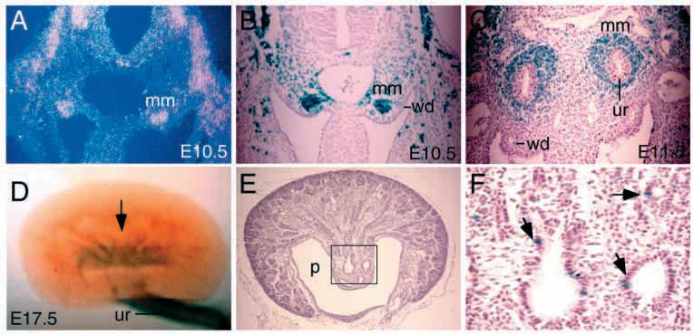
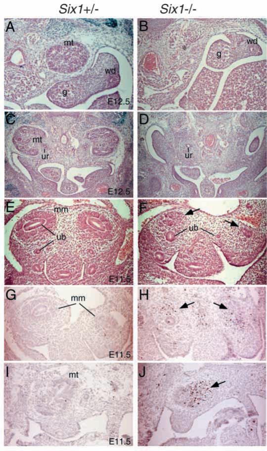
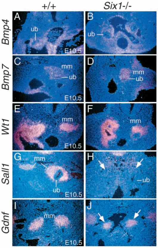
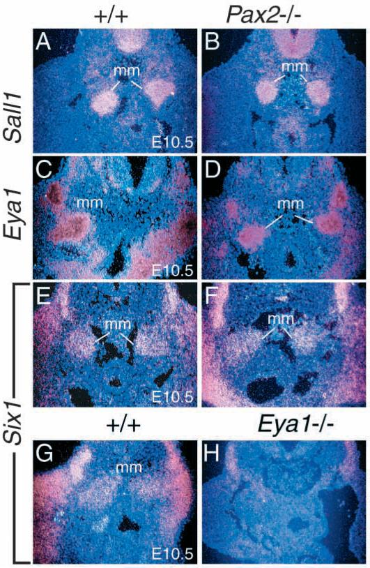
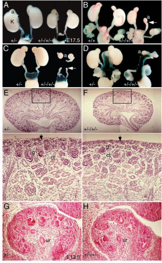
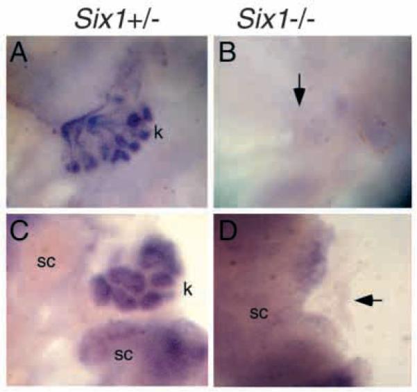
The murine Six gene family, homologous to Drosophila sine oculis (so) which encodes a homeodomain transcription factor, is composed of six members (Six1-6). Among the six members, only the Six2 gene has been previously shown to be expressed early in kidney development, but its function is unknown. We have recently found that the Six1 gene is also expressed in the kidney. In the developing kidney, Six1 is expressed in the uninduced metanephric mesenchyme at E10.5 and in the induced mesenchyme around the ureteric bud at E11.5. At E17.5 to P0, Six1 expression became restricted to a subpopulation of collecting tubule epithelial cells. To study its in vivo function, we have recently generated Six1 mutant mice. Loss of Six1 leads to a failure of ureteric bud invasion into the mesenchyme and subsequent apoptosis of the mesenchyme. These results indicate that Six1 plays an essential role in early kidney development. In Six1(-/-) kidney development, we have found that Pax2, Six2 and Sall1 expression was markedly reduced in the metanephric mesenchyme at E10.5, indicating that Six1 is required for the expression of these genes in the metanephric mesenchyme. In contrast, Eya1 expression was unaffected in Six1(-/-) metanephric mesenchyme at E10.5, indicating that Eya1 may function upstream of Six1. Moreover, our results show that both Eya1 and Six1 expression in the metanephric mesenchyme is preserved in Pax2(-/-) embryos at E10.5, further indicating that Pax2 functions downstream of Eya1 and Six1 in the metanephric mesenchyme. Thus, the epistatic relationship between Pax, Eya and Six genes in the metanephric mesenchyme during early kidney development is distinct from a genetic pathway elucidated in the Drosophila eye imaginal disc. Finally, our results show that Eya1 and Six1 genetically interact during mammalian kidney development, because most compound heterozygous embryos show hypoplastic kidneys. These analyses establish a role for Six1 in the initial inductive step for metanephric development.


Figures

Fig. 1
Fig. 1
Expression of Six1 during kidney development analyzed by in situ and X-gal staining of heterozygous Six1lacZ embryos for the Six1lacZ allele. (A) In situ hybridization showing Six1 expression in metanephric mesenchyme (mm) at E10.5. (B) X-gal staining of Six1lacZ heterozygous embryos showing strong Six1lacZ expression at E10.5, similar to that observed by in situ hybridization (A). (C) At E11.5, Six1 is expressed in the induced mesenchyme (mm) around the ureteric bud epithelium (ur). (D) X-gal staining of E17.5 Six1lacZ heterozygous kidneys showing Six1lacZ expression in collecting tubules (arrow). Six1lacZ is also expressed in the muscles surrounding the ureter (ur). (E,F) Transverse sections of X-gal-stained Six1lacZ heterozygous kidneys at E17.5 revealed that the Six1lacZ-expressing cells are localized in the collecting tubules (arrows). F is higher magnification of the boxed area in E. p, renal pelvis; wd, Wolffian duct.
Fig. 2
Fig. 2
Kidney development in Six1-deficient mice. (A-D) Metanephros (mt) in Six1+/− (A,C) and Six1−/− (B,D) mice at E12.5. Kidneys are absent in Six1−/− mice (*), but ureter (ur) was observed in the left primordium (D). The genital tracts appeared to be normal both in males and females. (E) In Six1+/− embryos of E11.5, ureteric bud (ub) grows out from Wolffian duct and invades the mesenchyme and the metanephric mesenchyme (mm) are condensed around the bulging ureteric bud. (F) In Six1−/− embryos, although the ureteric buds grow out from Wolffian ducts, they fail to invade the mesenchyme (arrows) completely. (G-J) TUNEL analysis of metanephros in Six1+/− and Six1−/− embryos at E11.5. Note the apoptotic cells were markedly increased in Six1−/− metanephric mesenchyme (arrows). g, gonad; wd, Wolffian duct.
Fig. 3
Fig. 3
Six1 is required for the expression of Pax2 and Six2 but not Eya1 in the metanephric mesenchyme (mm) at E10.5. (A-D) Pax2 is normally expressed in the intermediate mesoderm (A,B), the metanephric mesenchyme before and after induction and in the ureteric epithelium (ub). In Six1−/− embryos, however, its expression in the metanephric mesenchyme at E10.5 is undetectable (arrows in D). (E,F) Eya1 is expressed in the metanephric mesenchyme before and after induction and its expression is not affected in Six1−/− mesenchyme at E10.5. (G,H) Six2 is also expressed in the metanephric mesenchyme before and after induction and its expression is undetectable in Six1−/− mesenchyme at E10.5 (arrows).
Fig. 4
Fig. 4
Six1 is required for the expression of Sall1 in the metanephric mesenchyme (mm). (A,B) Bmp4 is normally expressed in the mesenchyme around the ureteric stalk (ub) and its expression was not affected in Six1−/− embryos at E10.5. (C,D) Bmp7 is normally expressed in the ureteric bud (ub) and metanephric mesenchyme at E10.5 and its expression levels are normal in both structures in Six1−/− embryos at E10.5. However, its expression domain is reduced in size in Six1−/− metanephric mesenchyme (D). (E,F) Wt1 is widely expressed in the mesenchyme and urogenital ridge region during kidney development and its expression level is not affected in Six1−/− embryos at E10.5. However, its expression domain in the metanephric mesenchyme also appears to be reduced in size in E10.5 Six1−/− embryos. (G,H) Sall1 is expressed in the metanephric mesenchyme before and after induction, however its expression was undetectable in the mesenchyme in Six1−/− embryos at E10.5 (arrows). (I,J) Gdnf is expressed in the metanephric mesenchyme and its expression levels are normal in Six1−/− embryos at E10.5. However, its expression domain appears to be reduced in size in Six1−/− embryos at E10.5 (arrows).
Fig. 5
Fig. 5
Sall1, Eya1 and Six1 expression is not affected in Pax2−/− metanephric mesenchyme (mm). (A,B) Sall1 is expressed in the metanephric mesenchyme and its expression is not affected in Pax2−/− mesenchyme at E10.5. (C,D) Eya1 is expressed in the metanephric mesenchyme and its expression level is unaffected in Pax2−/− embryos at E10.5. (E,F) Six1 is expressed in the metanephric mesenchyme and its expression level is also unaffected in Pax2−/− embryos at E10.5. (G,H) However, Six1 expression in Eya1−/− mesenchyme is significantly reduced when compared to that in wild-type embryos at E10.5. Its expression in the limb bud and somites is also significantly reduced. Six homozygous embryos were used for each probe.
Fig. 6
Fig. 6
Renal hypoplasia or agenesis in Eya1/Six1 double heterozygous animals. (A-D) E17.5 wild type, Six1+/− or Eya1+/−/Six1+/− kidneys. Samples shown in A, C and D were X-gal stained for Six1lacZ and it is expressed in the testis (t) and muscles surrounding the ureters and kidneys (D). Eya1+/−/Six1+/− animals show either smaller kidneys bilaterally (A), a small kidney on one side and no kidney on the other side (B), or complete absence of kidneys (C,D). In some Eya1+/−/Six1+/− animals that show renal agenesis, ureters that end blindly are observed (arrow in B,C). Adrenal glands and the genital tracts appeared to be normal in all compound heterozygous animals analyzed so far. (E,F) Histological analysis of kidneys of E17.5 Six1+/− and Eya1+/−/Six1+/− animals. The number of nephrons was markedly reduced in the double heterozygous kidneys, but normal developing structures are present. The lower panels are higher magnification of the boxed areas. In Six1+/− animals, the differentiating metanephric cap tissue (vesicles) in the peripheral nephrogenic zone, in which ureteric bud branching and induction of new nephrons takes place, are morphologically apparent (arrow in E). However, in the hypoplastic Eya1+/−/Six1+/− kidney, the differentiating metanephric vesicles in the peripheral nephrogenic zone were largely reduced in number (arrow in F). (G,H) A reduced number of ureteric bud branches is detected in the compound heterozygous animals at E13.5. ct, convoluting tubule; g, glomerulus; ur, ureter.
Fig. 7
Fig. 7
Six1 mutant mesenchyme is unresponsive to induction. (A) E11.0 Six1+/− kidney rudiments cultured for 5 days and stained with Pax2 in situ probe. After 5 days of culture, they developed into a fully branched kidney structure (k) showing Pax2 expression in the collecting tubules and in the nephrons. (B) E11.0 Six1−/− metanephric rudiments cultured for 5 days and stained with Pax2 in situ probe. No kidney formation was observed (arrow). (C) E11.0 Six1+/− metanephric mesenchyme cultured with spinal cord (sc) for 5 days and stained with Pax2 in situ probe. The Six1+/− mesenchymal cultures exhibited characteristic tubules (k) showing Pax2 expression. (D) E11.0 Six1−/− metanephric mesenchyme cultured with hetorozygous spinal cord for 5 days and stained with Pax2 in situ probe. None of the Six1−/− mesenchymes exhibited any sign of tubule formation. Note the disappearance of Six1 mutant mesenchyme (arrow), which shows no Pax2 expression.

References

    1. Al-Awqati Q, Oliver JA. Stem cells in the kidney. Kidney Int. 2002;61:387–395. - PubMed
    1. Ashley DJB, Mostofi FK. Renal agenesis and dysgenesis. J. Urol. 1960;83:211–230. - PubMed
    1. Brophy PD, Ostrom L, Lang KM, Dressler GR. Regulation of ureteric bud outgrowth by Pax2-dependent activation of the glial derived neurotrophic factor gene. Development. 2001;128:4747–4756. - PubMed
    1. Buller C, Xu X, Marquis V, Schwanke R, Xu P-X. Molecular effects of Eya1 domain mutations causing organ defects in BOR syndrome. Hum. Mol. Genet. 2001;10:2775–2781. - PubMed
    1. Cacalano G, Farinas I, Wang LC, Hagler K, Forgie A, Moore M, Armanini M, Phillips H, Ryan AM, Reichardt LF, et al. GFRalpha1 is an essential receptor component for GDNF in the developing nervous system and kidney. Neuron. 1998;21:53–62. - PMC - PubMed

Publication types

MeSH terms