

doi: 10.1101/gad.1092303.
Epub 2003 Jun 3.
Periodic repression by the bHLH factor Hes7 is an essential mechanism for the somite segmentation clock
Affiliations
- PMID: 12783854
- PMCID: PMC196074
- DOI: 10.1101/gad.1092303
Item in Clipboard
Periodic repression by the bHLH factor Hes7 is an essential mechanism for the somite segmentation clock
Genes Dev.
.
Abstract
Hes7, a bHLH gene essential for somitogenesis, displays cyclic expression of mRNA in the presomitic mesoderm (PSM). Here, we show that Hes7 protein is also expressed in a dynamic manner, which depends on proteasome-mediated degradation. Spatial comparison revealed that Hes7 and Lunatic fringe (Lfng) transcription occurs in the Hes7 protein-negative domains. Furthermore, Hes7 and Lfng transcription is constitutively up-regulated in the absence of Hes7 protein and down-regulated by stabilization of Hes7 protein. Thus, periodic repression by Hes7 protein is critical for the cyclic transcription of Hes7 and Lfng, and this negative feedback represents a molecular basis for the segmentation clock.
Figures
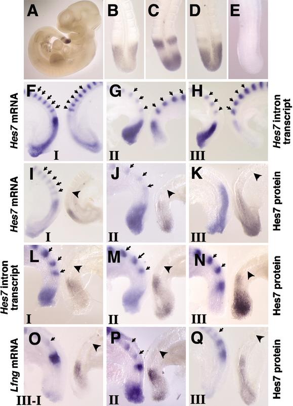
Spatial relationship of Hes7 protein, Hes7 mRNA, Hes7 nascent transcript, and Lfng mRNA in the PSM. (A–E) The expression pattern of Hes7 protein was examined by whole-mount immunochemistry. Hes7 immunoreactivity is specifically observed in the PSM of wild-type embryos (A). At E10.5, wild-type embryos display various expression patterns (B, n = 8; C, n = 5; D, n = 8). No signal is detected in Hes7-null mice (E, n = 3). (F–H) Comparison of the expression of Hes7 mRNA (left) and Hes7 nascent transcript (right) in the bisected caudal portions of E9.5 embryos. The latter is detected by the Hes7 intron probe. (I–Q) The expression of Hes7 protein (right) was compared with the expression of Hes7 mRNA (I–K, left), Hes7 nascent transcript (L–N, left), and Lfng mRNA (O–Q, left). The expression patterns are categorized into three phases: phase I (I,L,O), phase II (J,M,P), and phase III (K,N,Q). Hes7 protein-positive domains and the regions for Hes7 nascent transcript and Lfng mRNA are mutually exclusive. The established somites are stained with the Uncx4.1 probe for spatial alignment (arrows). The boundary between S0 and S-I is indicated by an arrowhead.
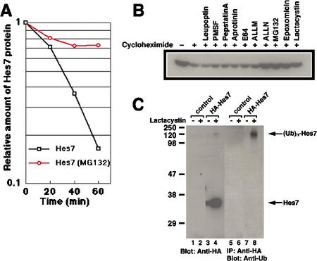
Hes7 protein is degraded by the ubiquitin–proteasome system. (A) C3H10T1/2 cells were transfected with the HA-Hes7 expression vector on the previous day. The cells were cultured in the presence of cycloheximide plus MG132 or cycloheximide only for 30 min and then harvested at the indicated time points. Whole-cell extracts were probed with anti-HA antibody, and the relative immunoreactive activities of the signals were measured. Hes7 protein is degraded with a half-life of 23.1 ± 8.7 min, whereas it is stabilized in the presence of the proteasome inhibitor MG132. (B) Cells were transfected with the HA-Hes7 expression vector on the previous day. The cells were cultured for 2 h with various inhibitors, as indicated above each lane, in the presence (+) of cycloheximide to inhibit new protein synthesis. Cell extracts were probed with anti-HA antibody. Note that Hes7 protein is stabilized by proteasome inhibitors (ALLN, MG132, epoxomicin, and lactacystin) but not by other protease inhibitors (leupeptin, PMSF, pepstatinA, aprotinin, E64, and ALLM). (C) C3H10T1/2 cells were transfected with the HA-Hes7 expression vector and cultured overnight in the presence (+) or absence (-) of lactacystin (20 μM). In the four left lanes, whole-cell extracts were probed with anti-HA antibody. In the presence of lactacystin, Hes7 protein is stabilized and higher-molecular-weight bands appear (lane 4). In the four right lanes, whole-cell extracts were immunoprecipitated with anti-HA antibody and probed with anti-ubiquitin antibody. The high-molecular-weight species are highly reactive to anti-ubiquitin antibody (lane 8).
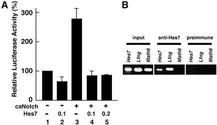
Analysis of Hes7 and Lfng promoters. (A) The luciferase vector under the control of the Hes7 promoter was cotransfected with or without the expression vector for caNotch and/or the expression vector for Hes7 (0.1 or 0.2μg). The activity of the luciferase vector with the Hes7 promoter alone (lane 1) is taken as 100%. Relative luciferase activities shown with a standard error are the average of three independent experiments performed in duplicate. Hes7 overrides the caNotch-induced activation of the Hes7 promoter (lanes 4,5). (B) ChIP analysis. Anti-Hes7 antibody specifically precipitates the chromatin containing the Hes7 and Lfng promoter regions, but not the Math6 promoter region, from PSM tissues. Preimmune serum does not precipitate these promoters.
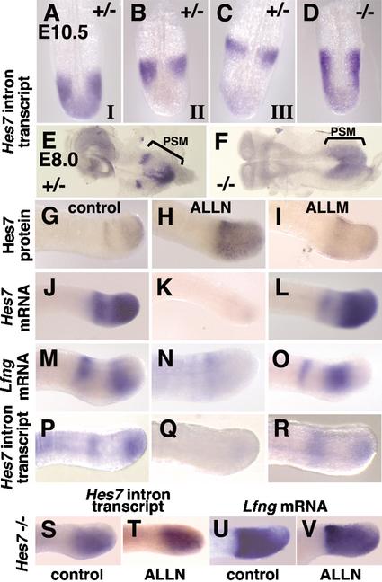
Dynamic expression of Hes7 mRNA is affected by Hes7 protein. (A–F) In situ hybridization was performed with the Hes7 intron probe, which detects the expression of the nascent transcript of both mutant and wild-type Hes7 alleles. The expression is dynamic in Hes7+/- embryos at E10.5 (A, phase I, n = 7; B, phase II, n = 7; C, phase III, n = 8) and E8.0 (E, n = 4). In contrast, the nascent transcript is expressed throughout the PSM in Hes7-null embryos both at E10.5 (D, n = 9) and at E8.0 (F, n = 4). (G–V) The caudal part of E10.5 embryos of wild type (G–R) and Hes7-/- (S–V) was cultured for 2 h with 100 μM ALLN, with 100 μM ALLM, or without protease inhibitors, as indicated. The expression patterns of Hes7 protein (G–I), Hes7 mRNA (J–L), Lfng mRNA (M–O,U,V), and Hes7 nascent transcript (P–R,S,T) were examined. Treatment of proteasome inhibitors stabilizes Hes7 protein (H) and down-regulates Hes7 and Lfng transcription (K,N,Q), compared with the control and ALLM-treated explants. In contrast, such repression does not occur in Hes7-/- PSM (S–V).
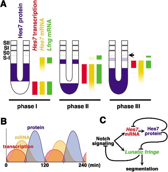
Summary of spatial and temporal expression patterns of Hes7 and model of the mechanism for the segmentation clock. (A) Spatial relationship of Hes7 protein, Hes7 transcription, Hes7 mRNA, and Lfng mRNA in the mouse PSM. Hes7 and Lfng transcription does not occur in the Hes7 protein-positive domains in all three phases. The prospective somite boundary at phase III is indicated by an arrow. (B) Temporal relationship of Hes7 protein, Hes7 mRNA, and Hes7 transcription in PSM cells. (C) A model of the mechanism for the segmentation clock based on the negative feedback loop of Hes7. Periodic repression by Hes7 protein is essential for cyclic expression and synchronization of Hes7 and Lfng mRNA.
References
-
- Aulehla A. and Johnson, R.L. 1999. Dynamic expression of Lunatic fringe suggests a link between notch signaling and an autonomous cellular oscillator driving somite segmentation. Dev. Biol. 207: 49–61. - PubMed
-
- Aulehla A., Wehrle, C., Brand-Saberi, B., Kemler, R., Gossler, A., Kanzler, B., and Herrmann, B.G. 2003. Wnt3a plays a major role in the segmentation clock controlling somitogenesis. Dev. Cell 4: 395–406. - PubMed
-
- Bessho Y., Miyoshi, G., Sakata, R., and Kageyama, R. 2001a. Hes7: A bHLH-type repressor gene regulated by Notch and expressed in the presomitic mesoderm. Genes Cells 6: 175–185. - PubMed
-
- Cole S.E., Levorse, J.M., Tilghman, S.M., and Vogt, T.F. 2002. Clock regulatory elements control cyclic expression of Lunatic fringe during somitogenesis. Dev. Cell 3: 75–84. - PubMed
Publication types
MeSH terms
Substances
LinkOut - more resources
Full Text Sources
Other Literature Sources
Molecular Biology Databases
Miscellaneous