

Density of neoplastic lymphoid infiltrate, CD8+ T cells, and CD1a+ dendritic cells in mycosis fungoides
- PMID: 12783973
- PMCID: PMC1769963
- DOI: 10.1136/jcp.56.6.453
Density of neoplastic lymphoid infiltrate, CD8+ T cells, and CD1a+ dendritic cells in mycosis fungoides
Abstract
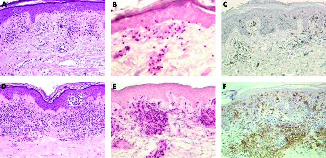
Background/aims: CD8+ T cells and epidermal/dermal dendritic cells expressing CD1a are found among neoplastic CD4+ T cells in mycosis fungoides (MF) lesions. This study analysed the relation of CD8+ tumour infiltrating lymphocytes (TILs), CD1a+ epidermal Langerhan's cells (LCs), and dermal dendritic cells (DDCs) to clinicopathological parameters in 46 MF cases.
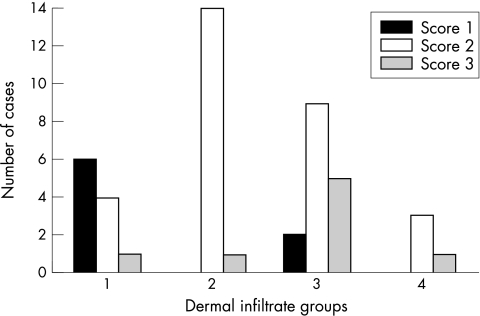
Methods: Pretreatment diagnostic biopsy specimens of 46 MF cases were submitted to histological analysis and immunohistochemistry. Four histological grades were defined based on the density of the neoplastic infiltrate: grade 1 (mild superficial perivascular infiltrate), grade 2 (moderate superficial perivascular infiltrate with some tendency to confluence), grade 3 (pronounced superficial band-like infiltrate), and grade 4 (deep nodular infiltrate). Epidermotropism was scored as low, moderate, or high. Numbers of CD8+ T cells and of dermal and epidermal CD1a+ cells were scored as 1 (low), 2 (moderate), and 3 (high). Correlations between these parameters and clinical data (age, sex, clinical type of lesions, stage, response to treatment, and recurrence) were analysed by the chi(2) test.
Results: Numbers of TILs and DDCs were associated with subepidermal infiltrates, being lower in less dense infiltrates, whereas there was no association between epidermal CD1a+ cells and the analysed parameters. Complete remission in treated patients was related to subepidermal infiltrates but not to TILs, LCs, or DDCs.
Conclusions: These results support the notion that CD8+ cells and dermal CD1a+ cells are active against tumour cells. MF with low numbers of TILs could represent an early stage of the disease, before TILs are activated against tumour specific antigens.
Figures

References
-
- Hansen ER. Immunoregulatory events in the skin of patients with cutaneous T-cell lymphoma. Arch Dermatol 1996;132:554–61. - PubMed
-
- Burg G, Dummer R, Haeffner A, et al. From inflammation to neoplasia: mycosis fungoides evolves from reactive inflammatory conditions (lymphoid infiltrates) transforming into neoplastic plaques and tumors. Arch Dermatol 2001;137:949–52. - PubMed
-
- Ralfkiaer E, Wantzin GL, Mason DY, et al. Phenotypic characterization of lymphocyte subsets in mycosis fungoides. Comparison with large plaque parapsoriasis and benign chronic dermatoses. Am J Clin Pathol 1985;84:610–19. - PubMed
-
- Ralfkiaer E. Immunohistological markers for the diagnosis of cutaneous lymphomas. Semin Diagn Pathol 1991;8:62–72. - PubMed
-
- Moll M, Reinhold U, Kukel S, et al. CD7-negative helper T cells accumulate in inflammatory skin lesions. J Invest Dermatol 1994;102:328–32. - PubMed
MeSH terms
Substances
LinkOut - more resources
Full Text Sources
Medical
Research Materials