

Histopathological characteristics of small diameter melanocytic naevi
- PMID: 12783974
- PMCID: PMC1769983
- DOI: 10.1136/jcp.56.6.459
Histopathological characteristics of small diameter melanocytic naevi
Abstract
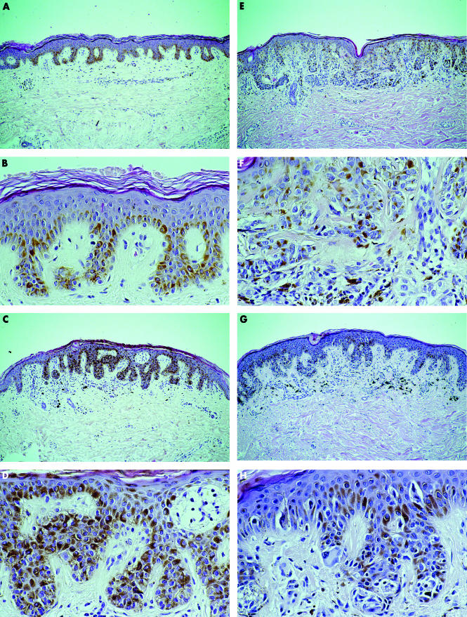
Background/aims: The clinical definition of an atypical naevus ("dysplastic naevus" or "naevus with architectural disorder and cytological atypia of melanocytes") stresses size larger than 5 mm in diameter as a major diagnostic criterion. Because malignant melanomas and their precursors may arise in smaller lesions, a histological study of melanocytic lesions smaller than 4 mm in diameter was conducted to evaluate their histological appearance.
Methods: Two hundred and sixty one naevi smaller than 4 mm in diameter were collected and characterised by histological examination into benign naevi without architectural disorder and naevi with architectural disorder and mild, moderate, and severe atypical melanocytes according to criteria used on larger lesions.
Results: Small melanocytic naevi covered the same complex histological spectrum from benign naevi to severely atypical naevi when compared with larger lesions. A high proportion of small naevi (72%) exhibited features diagnostic for naevi with architectural disorder and cytological atypia.
Conclusion: There is a discrepancy between histological and clinically defined atypical naevi. The same generally accepted criteria for the histological diagnosis of atypical naevi should be used for small melanocytic naevi in addition to large ones. Thus, small naevi exhibiting atypical features on histological examination should be categorised as atypical naevi, regardless of their small diameter.
Figures
Similar articles
-
Benign atypical naevi: diagnostic difficulties and continued controversy.Histopathology. 1999 Mar;34(3):189-98. doi: 10.1046/j.1365-2559.1999.00672.x. Histopathology. 1999. PMID: 10217558 Review.
-
Proposed alternative terminology and subclassification of so called "dysplastic naevi".J Clin Pathol. 1986 Feb;39(2):189-94. doi: 10.1136/jcp.39.2.189. J Clin Pathol. 1986. PMID: 3950040 Free PMC article.
-
The ultrastructure of dysplastic naevi: comparison with superficial spreading melanoma and common naevocellular naevi.Arch Dermatol Res. 1990;282(6):353-62. doi: 10.1007/BF00372084. Arch Dermatol Res. 1990. PMID: 2260880
-
My approach to atypical melanocytic lesions.J Clin Pathol. 2004 Nov;57(11):1121-31. doi: 10.1136/jcp.2003.008516. J Clin Pathol. 2004. PMID: 15509670 Free PMC article. Review.
-
The role of pattern analysis and the ABCD rule of dermoscopy in the detection of histological atypia in melanocytic naevi.Br J Dermatol. 2000 Aug;143(2):290-7. doi: 10.1046/j.1365-2133.2000.03653.x. Br J Dermatol. 2000. PMID: 10951135
Cited by
-
Histopathological diagnosis of small melanocytic lesions suspicious for malignant melanoma.An Bras Dermatol. 2017 May-Jun;92(3):375-378. doi: 10.1590/abd1806-4841.20175169. An Bras Dermatol. 2017. PMID: 29186251 Free PMC article.
-
Melanocytic lesions ≤ 6mm: Prospective series of 481 melanocytic trunk and limb lesions in Brazil.PLoS One. 2021 Jun 8;16(6):e0252162. doi: 10.1371/journal.pone.0252162. eCollection 2021. PLoS One. 2021. PMID: 34101726 Free PMC article.
References
-
- Garbe C, Büttner P, Wieβ J, et al. Risk factors for developing cutaneous melanoma and criteria for identifying persons at risk: multicenter case–control study of the central malignant melanoma registry of the German Dermatological Society. J Invest Dermatol 1996:102:695–9. - PubMed
-
- Grob JJ, Gouvernet J, Aymar D, et al. Count of benign melanocytic nevi as a major indicator of risk for nonfamilial nodular and superficial spreading melanoma. Cancer 1990;66:387–95. - PubMed
-
- Schneider JS, Moore DH, Sagebiel RW. Risk factors for melanoma incidence in prospective follow-up. The importance of atypical (dysplastic) nevi. Arch Dermatol 1994;103:202–5. - PubMed
-
- Snels DG, Hille ET, Gruis NA, et al. Risk of cutaneous malignant melanoma in patients with nonfamilial atypical nevi from a pigmented lesions clinic. J Am Acad Dermatol 1999;40:686–93. - PubMed
MeSH terms
LinkOut - more resources
Full Text Sources
Other Literature Sources
Medical