. 2003 Jun;48(5):1171-82.
doi: 10.1046/j.1365-2958.2003.03511.x.

The Escherichia coli amidase AmiC is a periplasmic septal ring component exported via the twin-arginine transport pathway

Affiliations

The Escherichia coli amidase AmiC is a periplasmic septal ring component exported via the twin-arginine transport pathway

Thomas G Bernhardt et al. Mol Microbiol. 2003 Jun.

Abstract

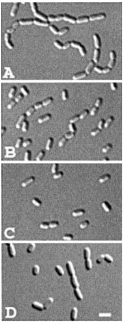
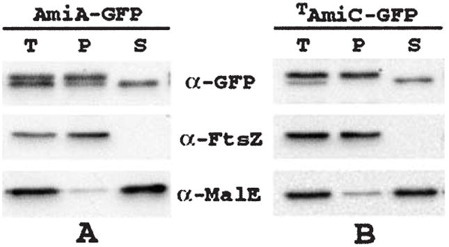
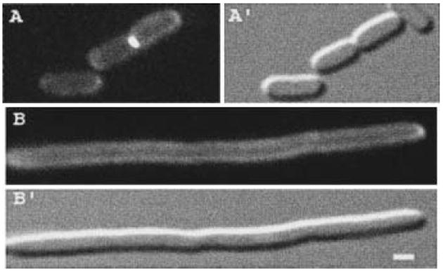
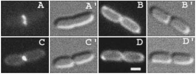
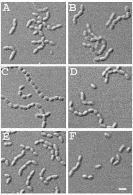
The N-acetylmuramoyl-l-alanine amidases of Escherichia coli (AmiA, B and C) are periplasmic enzymes that remove murein cross-links by cleaving the peptide moiety from N-acetylmuramic acid. Ami- cells form chains, indicating that the amidases help to split the septal murein. Interestingly, cells defective in the twin-arginine protein transport (Tat) pathway show a similar division defect. We find that both AmiA and AmiC are routed to the periplasm via Tat, providing an explanation for the Tat- division phenotype. Taking advantage of the ability of Tat to export prefolded (fluorescent) green fluorescent protein (GFP) to the periplasm, we sublocalized AmiA and AmiC in live cells using functional fusions to GFP. Interestingly, the periplasmic localization of the fusions differed markedly. AmiA-GFP appeared to be dispersed throughout the periplasm in all cells. AmiC-GFP similarly appeared throughout the periplasm in small cells, but was concentrated almost exclusively at the septal ring in constricting cells. Recruitment of AmiC to the ring was mediated by an N-terminal non-amidase targeting domain and required the septal ring component FtsN. AmiC therefore replaces FtsN as the latest known recruit to the septal ring and is the first entirely periplasmic component to be localized.


Figures

Fig. 1
Fig. 1
Amidase domains and plasmid and phage inserts. A. Signal sequences of E. coli periplasmic murein amidases AmiA, B and C (SWISSPROT accession P36548, P26365 and Q46929), as predicted by signalp version 1.1 (Nielsen et al., 1997). Potential Tat-targeting motifs (Berks et al., 2000; DeLisa et al., 2002) in AmiA and AmiC are underlined. B–D. Domain organization of AmiA (B), AmiC (C) and AmiB (D), plasmid and phage inserts and summary of functionality and sublocalization studies. Predicted signal sequences (SS), septal targeting (T) and catalytic (C) domains, relevant residue numbers and the approximate position of a native amiC promoter are indicated in the diagrams. Plasmid and phage inserts (solid lines), the lac promoter (arrows), gfpmut2 (boxes) and the presence of a non-native ribosome binding site (*) are indicated below the diagrams. The amidase domains were drawn based on the Pfam database (Bateman et al., 2002). Columns on the right-hand side summarize the functionality and sublocalization studies. TB53 [ΔamiA ΔamiC] cells carrying the indicated construct were grown in the presence of 0, 50, 100, 250 and 500 μM IPTG, and their division phenotypes (Phen.) were examined. ccc, severe chaining phenotype, over 80% of cells in chains of 4–14 units; c, mild chaining phenotype, 10–30% of cells in chains of 4–6 units; wt, wild type, less than 5% of cells in chains. Minimum IPTG concentrations (in μM) required to attain the indicated phenotype were: 0 (pTB27, λTB27, pTB39), 50 (pTB28, pTB41), 100 (λTB28), 250 (pTB32) and 500 (pTB33). Plasmids pTB34 and pTB37 failed to suppress chaining at all concentrations. The localization patterns (Loc.) of the fusion proteins are given in the second column: P, peripheral in all cells; P/R, accumulated at the septal ring in constricting cells, peripheral otherwise; ?, cytoplasmic fluorescence but localization unreliable because of excessive degradation of the fusion protein.
Fig. 2
Fig. 2
Functionality of the amidase fusion proteins. Differential interference contrast (DIC) micrographs show representative fields of TB53/pMLB1113 [ΔamiA ΔamiC/vector] (A), TB53/pTB32 [ΔamiA ΔamiC/PlacamiA–gfp] (B), TB53(λTB27) [ΔamiA ΔamiC (Plac∷PamiCamiC–gfp)] (C) and TB53/pTB33 [ΔamiA ΔamiC/PlacamiB–gfp] cells grown overnight in LB with no (C), 250 μM (A and B) or 500 μM (D) IPTG. Bar equals 2 μm.
Fig. 3
Fig. 3
Immunoblot analyses of GFP fusions. Blots probed with anti-GFP antibodies. Lanes in (A) contained whole-cell extracts of MC4100/pTB32 [wt/PlacamiA–gfp] (1) and B1LKO/pTB32 [ΔtatC/PlacamiA–gfp] (2). Cells were grown with 250 μM IPTG. The arrow indicates the positions of processed and unprocessed forms of AmiA–GFP (a). Lanes in (B) contained extracts of MC4100/pTB28 [wt/PlacamiC–gfp] (1), B1LKO/pTB28 [ΔtatC/PlacamiC–gfp] (2), MC4100/pTB34 [wt/PlacTamiC–gfp] (3), B1LKO/pTB34 [ΔtatC/PlacTamiC–gfp] (4) and TB54/pTB28 [PBADftsN/PlacamiC–gfp] (5 and 6). Cells were grown with 50 μM (1–4) or 100 μM (5 and 6) IPTG, and without (1–4 and 6) or with (5) 0.2% arabinose. Arrows indicate the positions of processed and unprocessed forms of AmiC–GFP (a) and TAmiC–GFP (b). Lanes in (C) contained extracts of TB28/pTB39 [wt/PlacamiC–gfp] (1) and TB28/pTB41 [wt/PlacCamiC–gfp] (2). Cells were grown with 10 μM IPTG. Arrows indicate the positions of processed and unprocessed forms of AmiC–GFP (a) and CAmiC–GFP (b). Note that the processed and unprocessed forms of the fusions to full-length AmiC migrated too closely to be distinguishable on these blots. For, this, as well as for Figs 5-8, cells were grown in M9-based medium to OD600 = 0.4–0.7.
Fig. 4
Fig. 4
Release of AmiA–GFP and TAmiC–GFP from spheroplasts. Strains TB28/pTB32 [wt/PlacamiA–gfp] (A) and TB28/pTB34 [wt/PlacTamiC–gfp] (B) were grown in LB supplemented with 0.2% maltose and 250 (A) or 50 (B) μM IPTG. One aliquot of cells was used to prepare a total-cell extract. The remaining cells were converted to spheroplasts and pelleted by centrifugation (see Experimental procedures). The resulting pellet (P) and supernatant (S) fractions, along with the total-cell extract (T), were analysed by SDS-PAGE and immunoblotting for GFP, FtsZ or MalE as indicated. FtsZ and MalE served as markers for the cytoplasm and periplasm respectively. Note that cells grown in rich medium, as used here for efficient spheroplast formation, show a ratio of unprocessed (non-periplasmic) to processed (periplasmic) forms of either fusion that is significantly greater than the ratio observed in cells grown in M9-based medium (see Fig. 3; results not shown).
Fig. 5
Fig. 5
Aberrant distribution of AmiA–GFP and AmiC–GFP in a ΔtatC mutant. Fluorescence (A–E) and DIC (A′–E′) micrographs show live cells of strains MC4100/pTB32 [wt/PlacamiA–gfp] (A), B1LKO/pTB32 [ΔtatC/PlacamiA–gfp] (B), MC4100/pTB28 [wt/PlacamiC–gfp] (C and D) and B1LKO/pTB28 [ΔtatC/PlacamiC–gfp] (E) grown with 250 μM (A and B) or 50 μM (C–E) IPTG. Note the chaining phenotype of Tat cells (B and E). We infer that the deeply constricted cell in (D) was about to separate, whereas the one in (C) was still finishing constriction (see text). Bar equals 1 μm.
Fig. 6
Fig. 6
Localization of AmiA–GFP and AmiC–GFP. Fluorescence (A–G) and DIC (A′–G′) micrographs show the distribution of AmiA–GFP (A and B) and AmiC–GFP (C–G) in live cells. Strains TB28/pTB32 [wt/PlacamiA–gfp] (A and B), TB36(λTB27) [ΔamiC(Plac∷PamiCamiC–gfp)] (C and D) and TB28/pTB28 [wt/PlacamiC–gfp] (E and G) were grown with 250 μM (A and B), no (C and D) or 50 μM (E–G) IPTG. The arrow in (G) points to a faint ring-like accumulation of AmiC–GFP in a cell at an early stage of constriction. Bar equals 1 μm.
Fig. 7
Fig. 7
Localization of AmiC–GFP in FtsN-depleted filaments. Fluorescence (A and B) and DIC (A′ and B′) micrographs show live cells of strain TB54/pTB28 [PBADftsN/PlacamiC–gfp] grown with 100 μM IPTG in the presence (A) or absence (B) of 0.2% arabinose. Bar equals 1 μm.
Fig. 8
Fig. 8
Localization of AmiC–GFP deletion derivatives. Fluorescence (A–D) and DIC (A′–D′) micrographs show live cells of strain TB28[wt] carrying pTB34 [PlacTamiC–gfp] (A), pTB37 [PlacSSamiC–gfp] (B), pTB39 [PlacamiC–gfp] (C) or pTB41 [PlacCamiC–gfp] (D) and grown with 10 μM (B–D) or 50 μM (A) IPTG. Bar equals 1 μm.
Fig. 9
Fig. 9
Overexpression of amiB–gfp suppresses the chaining phenotype of Tat cells. DIC micrographs show representative fields of B1LKO/pTB32 [ΔtatC/PlacamiA–gfp] (A and B), B1LKO/pTB28 [ΔtatC/PlacamiC–gfp] (C and D) and B1LKO/pTB33 [ΔtatC/PlacamiB–gfp] (E and F) cells grown overnight in LB with no (A, C and E), 0.5 mM (F) or 1 mM (B and D) IPTG. Bar equals 2 μm.

References

    1. Adam M, Fraipont C, Rhazi N, Nguyen-Disteche M, Lakaye B, Frere JM, et al. The bimodular G57–V577 polypeptide chain of the class B penicillin-binding protein 3 of Escherichia coli catalyzes peptide bond formation from thiolesters and does not catalyze glycan chain polymerization from the lipid II intermediate. J Bacteriol. 1997;179:6005–6009. - PMC - PubMed
    1. Addinall SG, Bi E, Lutkenhaus J. FtsZ ring formation in fts mutants. J Bacteriol. 1996;178:3877–3884. - PMC - PubMed
    1. Addinall SG, Cao C, Lutkenhaus J. FtsN, a late recruit to the septum in Escherichia coli. Mol Microbiol. 1997;25:303–309. - PubMed
    1. Baba T, Schneewind O. Targeting of muralytic enzymes to the cell division site of Gram-positive bacteria: repeat domains direct autolysin to the equatorial surface ring of Staphylococcus aureus. EMBO J. 1998;17:4639–4646. - PMC - PubMed
    1. Bateman A, Birney E, Cerruti L, Durbin R, Etwiller L, Eddy SR, et al. The Pfam protein families database. Nucleic Acids Res. 2002;30:276–280. - PMC - PubMed

Publication types

MeSH terms

Substances