. 2003 Jun;69(6):3435-47.
doi: 10.1128/AEM.69.6.3435-3447.2003.

Use of PCR for direct detection of Campylobacter species in bovine feces

Affiliations

Use of PCR for direct detection of Campylobacter species in bovine feces

G Douglas Inglis et al. Appl Environ Microbiol. 2003 Jun.

Abstract

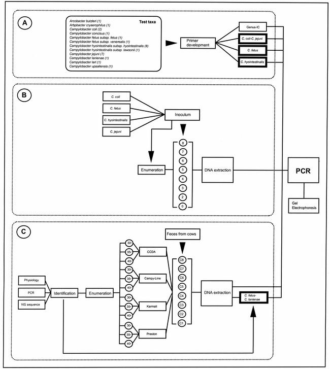
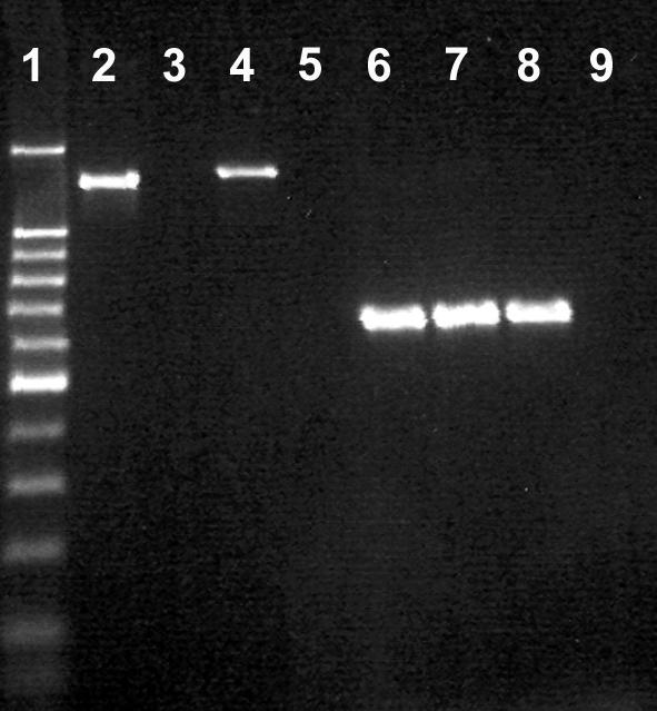
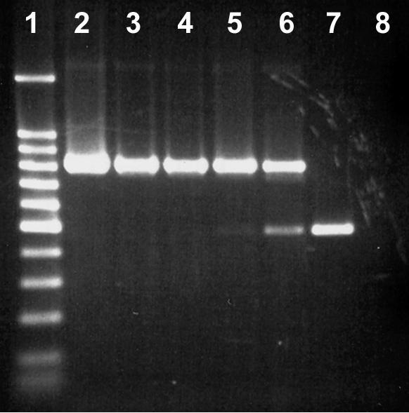
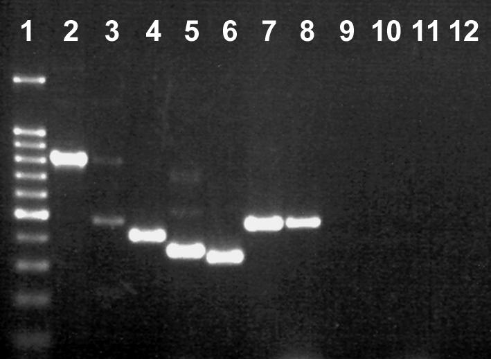
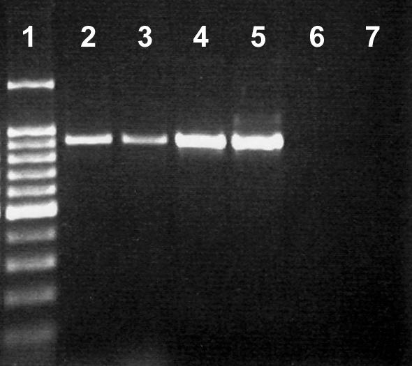
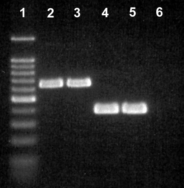
This study reports on the use of PCR to directly detect and distinguish Campylobacter species in bovine feces without enrichment. Inhibitors present in feces are a major obstacle to using PCR to detect microorganisms. The QIAamp DNA stool minikit was found to be an efficacious extraction method, as determined by the positive amplification of internal control DNA added to bovine feces before extraction. With nested or seminested multiplex PCR, Campylobacter coli, C. fetus, C. hyointestinalis, and C. jejuni were detected in all fecal samples inoculated at approximately 10(4) CFU g(-1), and 50 to 83% of the samples inoculated at approximately 10(3) CFU g(-1) were positive. At approximately 10(2) CFU g(-1), C. fetus, C. hyointestinalis, and C. jejuni (17 to 50% of the samples) but not C. coli were detected by PCR. From uninoculated bovine feces, a total of 198 arbitrarily selected isolates of Campylobacter were recovered on four commonly used isolation media incubated at three temperatures. The most frequently isolated taxa were C. jejuni (152 isolates) and C. lanienae (42 isolates), but isolates of C. fetus subsp. fetus, Arcobacter butzleri, and A. skirrowii also were recovered (</=2 isolates per taxon). Considerable variability was observed in the frequency of isolation of campylobacters among the four media and three incubation temperatures tested. With genus-specific primers, Campylobacter DNA was detected in 75% of the fecal samples, representing an 8% increase in sensitivity relative to that obtained with microbiological isolation across the four media and three incubation temperatures tested. With nested primers, C. jejuni and C. lanienae were detected in 25 and 67% of the samples, respectively. In no instance was DNA from either C. coli, C. fetus, or C. hyointestinalis detected in uninoculated bovine feces. PCR was more sensitive than isolation on microbiological media for detecting C. lanienae (17%) but not C. jejuni. Campylobacters are a diverse and fastidious group of bacteria, and the development of direct PCR not only will increase the understanding of Campylobacter species diversity and their frequency of occurrence in feces but also will enhance the knowledge of their role in the gastrointestinal tract of livestock and of the factors that influence shedding.


Figures

FIG. 1.
FIG. 1.
Flow diagram of the experiments conducted in each objective. (A) In the first objective, an internal control (IC) and primers for the genus Campylobacter, C. coli, C. jejuni, C. fetus, and C. hyointestinalis were developed. The presentation of two taxon names in the same box (on the right) represents multiplex reactions. Boxes with thick walls represent nested PCR. Names in the box labeled “Test taxa” represent taxa that were used to determine the specificities of PCRs; numbers in parentheses following the taxon names represent the numbers of isolates tested. (B) In the second objective, bovine feces were inoculated with various concentrations of C. coli, C. fetus, C. hyointestinalis, and C. jejuni (target densities in log10 CFU per gram are presented in circles); the control treatment (“0”) was not inoculated with campylobacters. CFUs were enumerated in the inoculated and control feces by using dilution plating. Concurrently, DNA was extracted from all fecal treatments and subjected to PCR with the primers shown in objective A, and amplicons were electrophoresed. (C) In the third objective, uninoculated bovine feces were collected from eight dairy cows (C1 to C8). Campylobacters were isolated and enumerated on four media maintained at three temperatures. Representative colonies were selected, established in pure cultures, and identified by using physiological characters, PCR with primers for specific taxa and, if required, extraction of DNA and sequencing of a portion of the 16S rRNA gene. During the course of this experiment, C. lanienae was frequently isolated on Karmali medium at 40°C. As a result, a nested primer set was developed for this taxon, and the C. lanienae primers were used in a multiplex PCR with primers for C. fetus. DNA was extracted from fecal samples obtained from the eight cows and subjected to PCR, and amplicons were electrophoresed. The experiments in the second and third objectives were conducted three times on separate occasions.
FIG. 2.
FIG. 2.
Comparison of the 16S (29) and 23S rDNA primers developed in the current study for detecting C. hyointestinalis. Lane 1, 100-bp molecular weight marker (the dark band was at 500 bp); lane 2, 16S primers with C. hyointestinalis subsp. hyointestinalis HBF; lane 3, 16S primers with C. hyointestinalis subsp. hyointestinalis HBE; lane 4, 16S primers with C. hyointestinalis subsp. hyointestinalis ATCC 35217; lane 5, 16S primers with C. hyointestinalis subsp. lawsonii NCTC 12901; lane 6, 23S primers with C. hyointestinalis subsp. hyointestinalis HBF; lane 7, 23S primers with C. hyointestinalis subsp. hyointestinalis HBE; lane 8, 23S primers with C. hyointestinalis subsp. hyointestinalis ATCC 35217; lane 9, 23S primers with C. hyointestinalis subsp. lawsonii NCTC 12901. An amplicon was not observed in either of the negative control reactions, and so these treatments were removed.
FIG. 3.
FIG. 3.
Impact of C. jejuni L102 genomic DNA concentration on the expression of the internal control amplicon. Lane 1, 100-bp molecular weight marker (the dark band was at 500 bp); lane 2, 10−1 dilution; lane 3, 10−2 dilution; lane 4, 10−3 dilution; lane 5, 10−4 dilution; lane 6, 10−5 dilution; lane 7, no template; lane 8, no internal control.
FIG. 4.
FIG. 4.
Multiplex PCR for detection of Campylobacter species in bovine feces. Lane 1, 100-bp molecular weight marker (the dark band was at 500 bp); lane 2, DNA from uninoculated feces amplified with the Campylobacter genus-specific primer set; lane 3, DNA from uninoculated feces amplified with the Campylobacter genus-specific primer set (note the weak genus amplicon and the internal control amplicon at 465 bp); lane 4, DNA from uninoculated feces amplified with the C. jejuni-C. coli multiplex nested primer set (note the C. jejuni amplicon at 413 bp); lane 5, DNA from uninoculated feces amplified with the C. fetus-C. lanienae nested primer set (note the C. lanienae amplicon at 360 bp); lane 6, DNA from feces inoculated with C. coli ATCC 49941 at a density of ≈103 CFU g−1 and amplified with the C. jejuni-C. coli multiplex nested primer set (note the C. coli amplicon at 330 bp); lane 7, DNA from feces inoculated with C. hyointestinalis subsp. hyointestinalis ATCC 35217 at a density of ≈102 CFU g−1 and amplified with the C. hyointestinalis seminested primer set (note the C. hyointestinalis amplicon at 468 bp); lane 8, DNA from feces inoculated with C. fetus ATCC 25936 at a density of ≈102 CFU g−1 and amplified with the C. fetus-C. lanienae nested primer set (note the C. fetus amplicon at 473 bp); lane 9, negative control for the internal control and Campylobacter genus-specific primers (no template added); lane 10, negative control for C. jejuni-C. coli multiplex primers; lane 11, negative control for C. fetus-C. lanienae multiplex primers; lane 12, negative control for C. hyointestinalis subsp. hyointestinalis primers.
FIG. 5.
FIG. 5.
Amplification of C. lanienae isolates with the 16S rRNA gene primers (31). Lane 1, 100-bp molecular weight marker (the dark band was at 500 bp); lane 2, C. lanienae L52; lane 3, C. lanienae L54; lane 4, C. lanienae NCTC 13004; lane 5, C. hyointestinalis subsp. lawsonii NCTC 12901; lane 6, C. hyointestinalis subsp. hyointestinalis ATCC 35217; lane 7, negative control.
FIG. 6.
FIG. 6.
Dendrogram based on a majority-rule consensus tree obtained from analyzing the partial 16S rRNA gene data set with the NEIGHBOR program (neighbor-joining option) and showing DNA sequence relatedness for campylobacters. The outgroup used in the analysis was C. jejuni, and isolates indicated by “T” represent type specimens. The bar represents 0.01 nucleotide substitution per base, and numbers at selected nodes indicate support obtained by bootstrap analysis (1,000 replicates) for the internal branches within the resulting trees.
FIG. 7.
FIG. 7.
Multiplex PCR of 16S and 23S rRNA genes for distinguishing Arcobacter species (19). Lane 1, 100-bp molecular weight marker ladder (the dark band was at 500 bp); lane 2, A. skirrowii L109 from bovine feces; lane 3, A. skirrowii L110 from bovine feces; lane 4, A. butzleri L111 from bovine feces; lane 5, A. butzleri ATCC 49616; lane 6, negative control.

References

    1. Atabay, H. I., and J. E. L. Corry. 1998. The isolation and prevalence of campylobacters from dairy cattle using a variety of methods. J. Appl. Microbiol. 84:733-740. - PubMed
    1. Ballagi-Pordany, A., and S. Belak. 1996. The use of mimics as internal standards to avoid false negatives in diagnostic PCR. Mol. Cell. Probes 10:159-164. - PubMed
    1. Bastyns, K., S. Chapelle, P. Vandamme, H. Goossens, and R. de Wachter. 1994. Species-specific detection of campylobacters important in veterinary medicine by PCR amplification of 23S rDNA areas. Syst. Appl. Microbiol. 17:563-568.
    1. Baylis, C. L., S. MacPhee, K. W. Martin, T. J. Humphrey, and R. P. Betts. 2000. Comparison of three enrichment media for the isolation of Campylobacter spp. from foods. J. Appl. Microbiol. 89:884-891. - PubMed
    1. Busato, A., D. Hofer, T. Lentze, C. Gaillard, and A. Burnens. 1999. Prevalence and infection risks of zoonotic enteropathogenic bacteria in Swiss cow-calf farms. Vet. Microbiol. 69:251-263. - PubMed

Publication types

Associated data

LinkOut - more resources