

doi: 10.1073/pnas.1230736100.
Epub 2003 Jun 3.
Germ-line transgenesis of the Tc1/mariner superfamily transposon Minos in Ciona intestinalis
Affiliations
- PMID: 12788975
- PMCID: PMC164655
- DOI: 10.1073/pnas.1230736100
Item in Clipboard
Germ-line transgenesis of the Tc1/mariner superfamily transposon Minos in Ciona intestinalis
Proc Natl Acad Sci U S A.
.
Abstract
The tadpole larva of the basal chordate Ciona intestinalis has the most simplified, basic body-plan of chordates. Because it has a compact genome with a complete draft sequence, a large quantity of EST/cDNA information, and a short generation time, Ciona is a suitable model for future genetics. We establish here a transgenic technique in Ciona that uses the Tc1/mariner superfamily transposon Minos. Minos was integrated efficiently into the genome of germ cells and transmitted stably to subsequent generations. In addition, an enhancer-trap line was obtained. This is a demonstration of efficient, Minos-mediated transgenesis in marine invertebrates.
Figures

Inheritance of GFP expression in Ciona F1 and F2 progeny. (A) Structure of a Minos construct, pMiLRCiTPO-gfp. The dotted line indicates the probe region used for Southern blot analysis shown in Fig. 2A. Arrows indicate the primer positions used for PCR analysis shown in Fig. 2B. LIR and RIR, left and right inverted repeats (IR) of Minos; pA, polyadenylation signal sequence; EI, EcoRI restriction site. (B–E) F1 progeny and the expression of GFP from Mi[CiTPOgfp]3 (B and C) and Mi[CiTPOgfp]2 (D and E). In C, GFP was seen at the anterior and posterior ends of the endostyle (red arrows). In E, endostyle (EN), peripharyngeal band (PB), dorsal tubercle (DT), and posterior margin of pharyngeal sac (PM) were marked by GFP. (F and G) GFP expression in F2 progeny derived from Mi[CiTPOgfp]3 (F) and Mi[CiTPOgfp]2 (G).
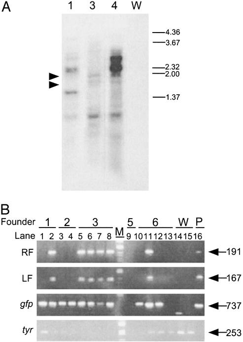
Genomic integration of Minos revealed by Southern blot and PCR analyses. (A) Genomic DNA isolated from F1 progeny of Mi[CiTPOgfp]1, 3, and 4 (lanes 1, 3, and 4) and wild type (lane W) were digested with EcoRI and electrophoresed. Right numbers indicate the sizes of markers (in kbp). There are several bands characteristic to each F1 progeny, in addition to similarly sized bands of ≈1.6 and 2.0 kbp (arrowheads). (B) Several F1 progeny lacked plasmid sequences of pMiLRCiTPO-gfp flanking Minos IR. Genomic DNA isolated from F1 progeny from Mi[CiTPOgfp]1–3, -5, -6 (–6), and wild type (W) were subjected to PCR analysis using primers described in Fig. 1 A. pMiLRCiTPO-gfp plasmid (P) was used as a positive control. Lane M indicates the marker. Right numbers show the size of bands (in bp). Note that several F1 (lanes 1, 3, 4, 10, and 12) lacked left flanking (LF) and right flanking (RF) sequences of IR of pMiLRCiTPO-gfp, although gfp was transmitted. PCR against tyrosinase gene (tyr) was done as an internal control.
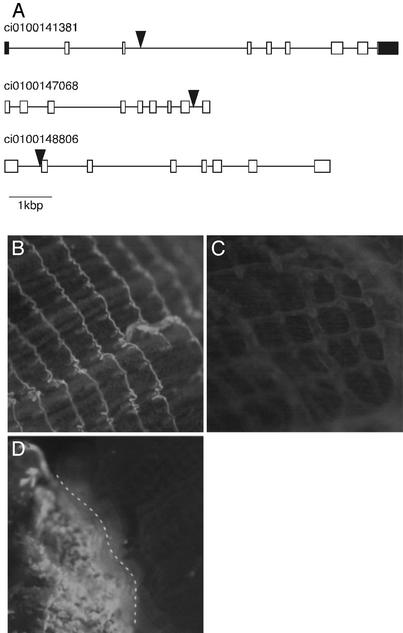
Examples of genes targeted by Minos and of enhancer-trap events. (A) Genes disrupted by Minos. Exons and introns are shown by boxes and lines, respectively. Exons corresponding to UTRs are labeled in black. Large triangles indicate the insertion sites of Minos. Grail numbers indicate the gene model in the C. intestinalis genome database (http://genome.jgi-psf.org/ciona4 ). (B–D) Two examples of enhancer-trap events. GFP was expressed in the pharyngeal gill of F2 progeny derived from Mi[CiTPOgfp]2 (B), but not in Mi[CiTPOgfp]3 (C). (D) A founder animal expressing GFP in the pharyngeal gill. Because the Minos insertion probably occurred in half of the precursor cells of the pharyngeal gill, only half of the gill expressed GFP (left side of the broken line).
Similar articles
-
High-throughput enhancer trap by remobilization of transposon Minos in Ciona intestinalis.Genesis. 2007 May;45(5):307-17. doi: 10.1002/dvg.20290. Genesis. 2007. PMID: 17464954
-
Enhancer detection in the ascidian Ciona intestinalis with transposase-expressing lines of Minos.Dev Dyn. 2008 Jan;237(1):39-50. doi: 10.1002/dvdy.21333. Dev Dyn. 2008. PMID: 17948255
-
Germline transgenesis of the chordate Ciona intestinalis with hyperactive variants of sleeping beauty transposable element.Dev Dyn. 2013 Jan;242(1):30-43. doi: 10.1002/dvdy.23891. Epub 2012 Nov 14. Dev Dyn. 2013. PMID: 23073965
-
Transposon mediated transgenesis in a marine invertebrate chordate: Ciona intestinalis.Genome Biol. 2007;8 Suppl 1(Suppl 1):S3. doi: 10.1186/gb-2007-8-s1-s3. Genome Biol. 2007. PMID: 18047695 Free PMC article. Review.
-
Germline transgenesis and insertional mutagenesis in the ascidian Ciona intestinalis.Dev Dyn. 2007 Jul;236(7):1758-67. doi: 10.1002/dvdy.21111. Dev Dyn. 2007. PMID: 17342755 Review.
Cited by
-
Stable germline transgenesis using the Minos Tc1/mariner element in the sea urchin Lytechinus pictus.Development. 2024 Oct 15;151(20):dev202991. doi: 10.1242/dev.202991. Epub 2024 Aug 19. Development. 2024. PMID: 39023164 Free PMC article.
-
Functional genetics for all: engineered nucleases, CRISPR and the gene editing revolution.Evodevo. 2014 Nov 18;5:43. doi: 10.1186/2041-9139-5-43. eCollection 2014. Evodevo. 2014. PMID: 25699168 Free PMC article. Review.
-
Maternal factor-mediated epigenetic gene silencing in the ascidian Ciona intestinalis.Mol Genet Genomics. 2010 Jan;283(1):99-110. doi: 10.1007/s00438-009-0500-4. Epub 2009 Nov 28. Mol Genet Genomics. 2010. PMID: 19946786
-
Transcriptional regulation of a horizontally transferred gene from bacterium to chordate.Proc Biol Sci. 2016 Dec 28;283(1845):20161712. doi: 10.1098/rspb.2016.1712. Proc Biol Sci. 2016. PMID: 28003446 Free PMC article.
-
The nervous system of the adult ascidian Ciona intestinalis Type A (Ciona robusta): Insights from transgenic animal models.PLoS One. 2017 Jun 26;12(6):e0180227. doi: 10.1371/journal.pone.0180227. eCollection 2017. PLoS One. 2017. PMID: 28651020 Free PMC article.
References
-
- Satoh, N. (1994) Developmental Biology of Ascidians (Cambridge Univ. Press, New York).
-
- Satoh, N. (2001) Differentiation (Berlin) 68, 1-12. - PubMed
-
- Corbo, J. C., Di Gregorio, A. & Levine, M. (2001) Cell 106, 535-538. - PubMed
-
- Conklin, E. G. (1905) J. Acad. Nat. Sci. 13, 1-119.
-
- Nishida, H. (1987) Dev. Biol. 121, 526-541. - PubMed
Publication types
MeSH terms
Substances
LinkOut - more resources
Full Text Sources
Research Materials
