. 2003 Jun 24;100(13):7847-52.
doi: 10.1073/pnas.1331360100. Epub 2003 Jun 3.

Maspin expression inhibits osteolysis, tumor growth, and angiogenesis in a model of prostate cancer bone metastasis

Affiliations

Maspin expression inhibits osteolysis, tumor growth, and angiogenesis in a model of prostate cancer bone metastasis

Michael L Cher et al. Proc Natl Acad Sci U S A. .

Abstract

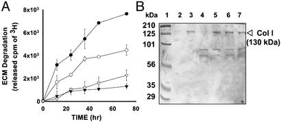
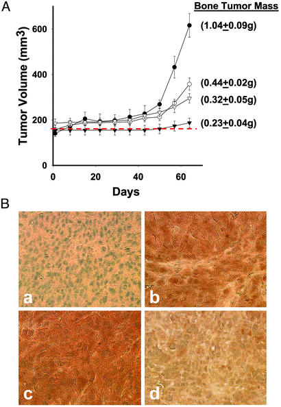
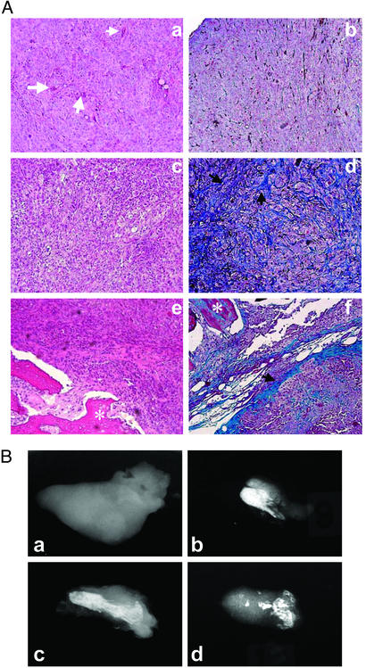
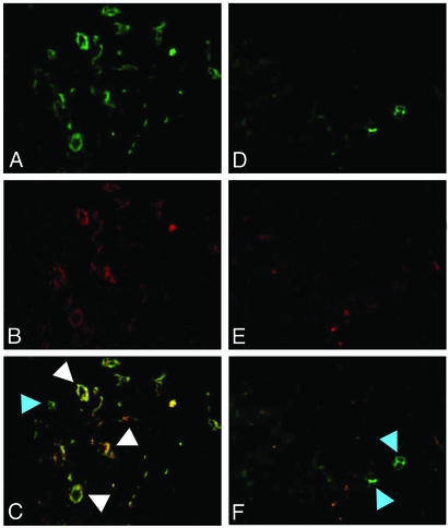
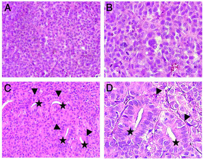
Emerging evidence indicates that tumor-associated proteolytic remodeling of bone matrix may underlie the capacity of tumor cells to colonize and survive in the bone microenvironment. Of particular importance, urokinase-type plasminogen activator (uPA) has been shown to correlate with human prostate cancer (PC) metastasis. The importance of this protease may be related to its ability to initiate a proteolytic cascade, leading to the activation of multiple proteases and growth factors. Previously, we showed that maspin, a serine protease inhibitor, specifically inhibits PC-associated uPA and PC cell invasion and motility in vitro. In this article, we showed that maspin-expressing transfectant cells derived from PC cell line DU145 were inhibited in in vitro extracellular matrix and collagen degradation assays. To test the effect of tumor-associated maspin on PC-induced bone matrix remodeling and tumor growth, we injected the maspin-transfected DU145 cells into human fetal bone fragments, which were previously implanted in immunodeficient mice. These studies showed that maspin expression decreased tumor growth, reduced osteolysis, and decreased angiogenesis. Furthermore, the maspin-expressing tumors contained significant fibrosis and collagen staining, and exhibited a more glandular organization. These data represent evidence that maspin inhibits PC-induced bone matrix remodeling and induces PC glandular redifferentiation. These results support our current working hypothesis that maspin exerts its tumor suppressive role, at least in part, by blocking the pericellular uPA system and suggest that maspin may offer an opportunity to improve therapeutic intervention of bone metastasis.


Figures

Fig. 1.
Fig. 1.
Analyses for ECM and hCol I degradation. (A) Degradation of metabolically labeled subendothelial 3H-ECM by the DU145-derived mock-transfected cells (•) and maspin-transfected clones M3 (○), M7 (▾), and M10 (▿). The radioactivity derived from 3H-ECM in the absence of PC cells was subtracted from the radioactivity of each sample at the corresponding time point. Data represent the average of two repeats. Each repeat was carried out in triplicate. Error bars = SD (n = 6). (B) Degradation of hCol I by the CM of mock-transfected cells and maspin-transfected clones 7 and 10 (lanes 4, 5, and 6, respectively). Proteins are revealed by Coomassie blue stain on SDS/PAGE. The molecular mass of intact hCol I is estimated as 130-kDa based on the molecular mass standards (lane1). Lanes 2 and 3 show hCol I incubated with HMW-uPA plus Glu-plasminogen and HMW-uPA alone, respectively. Lane 7 shows hCol I-incubated aprotinin in the CM of mock-transfected cells.
Fig. 2.
Fig. 2.
The effect of maspin on intraosseous tumor growth and uPA expression. (A) Bone tumor growth curves and end point tumor masses in an intraosseous SCID-human model. The PC bone tumors were derived from the mock-transfected control cells (•) and maspin-transfected clones M3, M7, and M10 (○, ▾, and ▿, respectively). The dashed line shows the average volume of the human bone implants before expansion by tumor cells. Each data point represents the average volume of PC bone tumors created from the indicated clonal cell line (n = 6), except for the mock-transfected DU145 cells, which had tumors due to the perioperative death of one animal (n = 5). Error bars = SEM. (B) Immunolocalization of Maspin and uPA. Immunohistochemistry was used to localize maspin protein in mock-transfected bone tumors (a) and maspin-transfectant bone tumors (b), as well as uPA in mock-transfectant bone tumors (c) and maspin-transfectant bone tumors (d). Brown indicates specific immunoreactivity, whereas the nuclei were counterstained blue. Representative images (×400) were acquired by using a SPOT digital camera (W. Nuhsbaum, McHenry, IL) interfaced with a Leica DM IRB microscope.
Fig. 3.
Fig. 3.
Histopathological and x-ray analyses of PC bone tumors. (A) Histopathological features of bone tumors. (a and b) The representative bone tumor of mock-transfected cells. (c–f) The representative bone tumors of maspin-transfected cells. a, d, and e are stained by hematoxylin/eosin and b, d, and f are stained by Masson's trichrome. (a–f, ×100). Black arrows indicate fibrosis, white arrows indicate microvessels, and white asterisks indicate remaining bone fragments. (B) X-ray imaging analyses of the PC bone tumors. The bright areas are residual mineralized bone and the gray areas indicate tumor mass without bone. (a) A representative bone tumor derived from the mock-transfected cells. (b–d) The representative bone tumors derived from maspin-transfected clones M3, M7, and M10, respectively.
Fig. 4.
Fig. 4.
Immunodetection of blood vessels in PC bone tumors. Blood vessel density in mock-transfectant tumors (A–C) and maspin-transfectant tumors (D–F) was monitored by immunofluorescent staining for factor VIII (green) and human CD 31 (red). The merged fluorescent images are shown in C (A and B) and F (D and E). All of the tissue sections were stained. Ten microscopic fields of each section were examined by using a Leica DM IRB fluorescence microscope. The fluorescent images were acquired by using a SPOT digital camera. (Magnification, ×100.)
Fig. 5.
Fig. 5.
Glandular morphology in maspin-transfectant PC bone tumors. (A and B) Mock-transfected tumor cells formed solid sheets with no glandular differentiation. (C and D) Many areas of maspin-transfectant bone tumors featured well formed small acini with lumena. A hematoxylin/eosin stain was used. (Magnification: A and C, ×200; B and D, ×400.) Arrows indicate glands with intact lumena, the tips of which are labeled with stars.

Similar articles

Cited by

References

    1. Tu, S. M., Millikan, R. E., Mengistu, B., Delpassand, E. S., Amato, R. J., Pagliaro, L. C., Daliani, D., Papandreou, C. N., Smith, T. L., Kim, J., et al. (2001) Lancet 357, 336-341. - PubMed
    1. Shimazaki, J., Higa, T., Akimoto, S., Masai, M. & Isaka, S. (1992) Adv. Exp. Med. Biol. 324, 269-275. - PubMed
    1. Akimoto, S., Furuya, Y., Akakura, K. & Ito, H. (1998) Endocr. J. 45, 97-104. - PubMed
    1. Revilla, M., Arribas, I., Sanchez, C. M., Villa, L. F., Bethencourt, F. & Rico, H. (1998) Prostate 35, 243-247. - PubMed
    1. Rubens, R. D. (1998) Eur. J. Cancer 34, 210-213. - PubMed

Publication types

MeSH terms