

Studies of epidemiology and seroprevalence of bovine noroviruses in Germany
- PMID: 12791840
- PMCID: PMC156573
- DOI: 10.1128/JCM.41.6.2300-2305.2003
Studies of epidemiology and seroprevalence of bovine noroviruses in Germany
Abstract
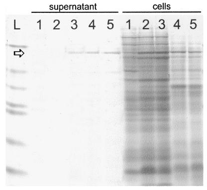
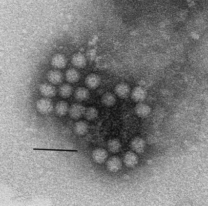
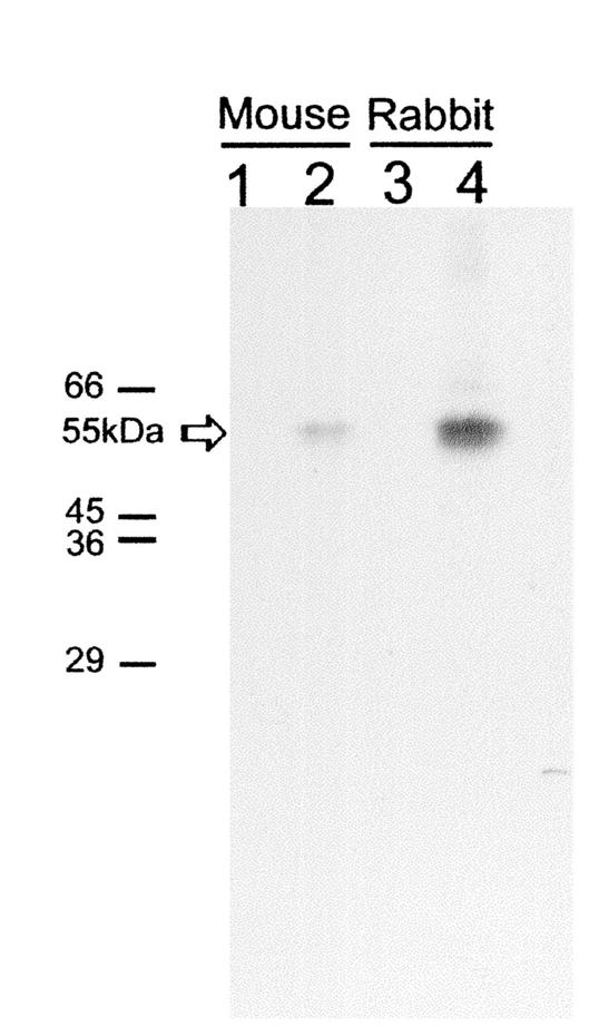
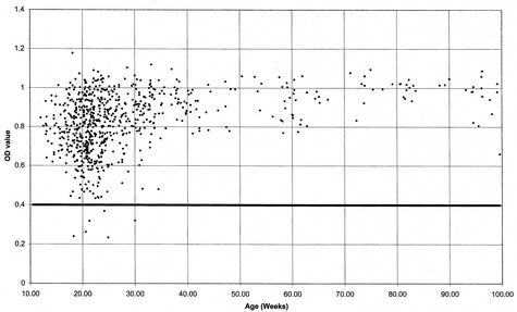
Jena virus (JV) is a bovine enteric calicivirus that causes diarrhea in calves. The virus is approximately 30 nm in diameter and has a surface morphology similar to the human Norwalk virus. The genome sequence of JV was recently described, and the virus has been assigned to the genus Norovirus of the family CALICIVIRIDAE: In the present study, the JV capsid gene encoded by open reading frame 2 was cloned into the baculovirus transfer vector pFastBac 1, and this was used to transform Escherichia coli to generate a recombinant bacmid. Transfection of insect cells with the recombinant baculovirus DNA resulted in expression of the JV capsid protein. The recombinant JV capsid protein undergoes self-assembly into virus-like particles (VLPs) similar to JV virions in size and appearance. JV VLPs were released into the cell culture supernatant, concentrated, and then purified by CsCl equilibrium gradient centrifugation. Purified JV VLPs were used to hyperimmunize laboratory animals. An antigen capture enzyme-linked immunosorbent assay (ELISA) was developed and characterized initially with clinical specimens containing defined human noroviruses and bovine diarrheal samples from calves experimentally infected with JV; the ELISA was specific only for JV. The ELISA was used to screen 381 diarrheal samples collected from dairy herds in Thuringia, Hesse, and Bavaria, Germany, from 1999 to 2002; 34 of these samples (8.9%) were positive for JV infection. The unexpectedly high prevalence of JV was confirmed in a seroepidemiological study using 824 serum or plasma samples screened using an anti-JV ELISA, which showed that 99.1% of cattle from Thuringia have antibodies to JV.
Figures
References
-
- Almeida, J. D., C. R. Craig, and T. E. Hall. 1978. Multiple viruses present in the faeces of a scouring calf. Vet. Rec. 102:170-171. - PubMed
-
- Bridger, J. C. 1990. Small viruses associated with gastroenteritis in animals, p. 161-182. In L. J. Saif and K. W. Theil (ed.), Viral diarrheas of man and animals. CRC Press, Boca Raton, Fla.
-
- Bridger, J. C., G. A. Hall, D. J. Reynolds, and J. F. Brown. 1983. Calici-like viruses in calf diarrhoea, p. 155-159. In Proceedings of the 4th International Symposium on Neonatal Diarrhoea. Vaccine & Infectious Disease Organization, Saskatoon, Saskatchewan, Canada.
-
- Chadwick, P. R., G. Beards, D. Brown, E. O. Caul, J. Cheesbrough, I. Clarke, A. Curry, S. O'Brien, K. Quigley, J. Sellwood, and D. Westmoreland. 2000. Management of hospital outbreaks of gastro-enteritis due to small round structured viruses. J. Hosp. Infect. 45:1-10. - PubMed
MeSH terms
Substances
LinkOut - more resources
Full Text Sources
Medical
