Species-specific identification of campylobacters by partial 16S rRNA gene sequencing
- PMID: 12791878
- PMCID: PMC156541
- DOI: 10.1128/JCM.41.6.2537-2546.2003
Species-specific identification of campylobacters by partial 16S rRNA gene sequencing
Abstract
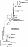
Species-specific identification of campylobacters is problematic, primarily due to the absence of suitable biochemical assays and the existence of atypical strains. 16S rRNA gene (16S rDNA)-based identification of bacteria offers a possible alternative when phenotypic tests fail. Therefore, we evaluated the reliability of 16S rDNA sequencing for the species-specific identification of campylobacters. Sequence analyses were performed by using almost 94% of the complete 16S rRNA genes of 135 phenotypically characterized Campylobacter strains, including all known taxa of this genus. It was shown that 16S rDNA analysis enables specific identification of most Campylobacter species. The exception was a lack of discrimination among the taxa Campylobacter jejuni and C. coli and atypical C. lari strains, which shared identical or nearly identical 16S rDNA sequences. Subsequently, it was investigated whether partial 16S rDNA sequences are sufficient to determine species identity. Sequence alignments led to the identification of four 16S rDNA regions with high degrees of interspecies variation but with highly conserved sequence patterns within the respective species. A simple protocol based on the analysis of these sequence patterns was developed, which enabled the unambiguous identification of the majority of Campylobacter species. We recommend 16S rDNA sequence analysis as an effective, rapid procedure for the specific identification of campylobacters.
Figures


Similar articles
-
Novel Campylobacter lari-like bacteria from humans and molluscs: description of Campylobacter peloridis sp. nov., Campylobacter lari subsp. concheus subsp. nov. and Campylobacter lari subsp. lari subsp. nov.Int J Syst Evol Microbiol. 2009 May;59(Pt 5):1126-32. doi: 10.1099/ijs.0.000851-0. Int J Syst Evol Microbiol. 2009. PMID: 19406805
-
Emerging thermotolerant Campylobacter species in healthy ruminants and swine.Foodborne Pathog Dis. 2011 Jul;8(7):807-13. doi: 10.1089/fpd.2010.0803. Epub 2011 Mar 27. Foodborne Pathog Dis. 2011. PMID: 21438765
-
Extensive 16S rRNA gene sequence diversity in Campylobacter hyointestinalis strains: taxonomic and applied implications.Int J Syst Bacteriol. 1999 Jul;49 Pt 3:1171-5. doi: 10.1099/00207713-49-3-1171. Int J Syst Bacteriol. 1999. PMID: 10425776
-
Detection and identification of Campylobacter spp. using the polymerase chain reaction.Cell Mol Biol (Noisy-le-grand). 1995 Jul;41(5):625-38. Cell Mol Biol (Noisy-le-grand). 1995. PMID: 7580843 Review.
-
Then and now: use of 16S rDNA gene sequencing for bacterial identification and discovery of novel bacteria in clinical microbiology laboratories.Clin Microbiol Infect. 2008 Oct;14(10):908-34. doi: 10.1111/j.1469-0691.2008.02070.x. Clin Microbiol Infect. 2008. PMID: 18828852 Review.
Cited by
-
Rapid detection of Campylobacter antigen by enzyme immunoassay leads to increased positivity rates.J Clin Microbiol. 2013 Feb;51(2):618-20. doi: 10.1128/JCM.02565-12. Epub 2012 Nov 21. J Clin Microbiol. 2013. PMID: 23175262 Free PMC article.
-
Oral Campylobacter species involved in extraoral abscess: a report of three cases.J Clin Microbiol. 2005 May;43(5):2513-5. doi: 10.1128/JCM.43.5.2513-2515.2005. J Clin Microbiol. 2005. PMID: 15872299 Free PMC article.
-
Campylobacter portucalensis sp. nov., a new species of Campylobacter isolated from the preputial mucosa of bulls.PLoS One. 2020 Jan 10;15(1):e0227500. doi: 10.1371/journal.pone.0227500. eCollection 2020. PLoS One. 2020. PMID: 31923228 Free PMC article.
-
Uncovering the boundaries of Campylobacter species through large-scale phylogenetic and nucleotide identity analyses.mSystems. 2024 Apr 16;9(4):e0121823. doi: 10.1128/msystems.01218-23. Epub 2024 Mar 26. mSystems. 2024. PMID: 38530055 Free PMC article.
-
A novel real-time PCR assay for quantitative detection of Campylobacter fetus based on ribosomal sequences.BMC Vet Res. 2016 Dec 15;12(1):286. doi: 10.1186/s12917-016-0913-3. BMC Vet Res. 2016. PMID: 27978826 Free PMC article.
References
-
- Bisping, W., and G. Amtsberg. 1988. Genus: Campylobacter, p. 215-231. In Colour atlas for the diagnosis of bacterial pathogens in animals. Paul Parey Scientific Publishers, Berlin, Germany.
-
- Blaser, M. J. 1997. Epidemiologic and clinical features of Campylobacter jejuni infections. J. Infect. Dis. 176:103-105. - PubMed
-
- Bolton, F. J., D. Coates, D. N. Hutchinson, and A. F. Godfree. 1987. A study of thermophilic campylobacters in a river system. J. Appl. Bacteriol. 62:167-176. - PubMed
Publication types
MeSH terms
Substances
LinkOut - more resources
Full Text Sources
Other Literature Sources
Medical
Molecular Biology Databases

